Abnormalities in a Newborn's Belly Button


Written and verified by the nurse Leidy Mora Molina
The umbilical cord is the structure that unites the baby with its mother during pregnancy and allows it to be nourished and oxygenated inside the uterus. At birth, this physical union is severed and later on, a scar will give way to the navel we all know. But did you know that some abnormalities in the newborn’s belly button can occur?
During the period of drying and falling off of the remaining stump of the cord, inconveniences can arise, some of which are potentially serious. For this reason, today we want to talk to you about abnormalities in a newborn’s belly button so that you recognize them and know how to act when you detect them.
1. Delayed detachment of the umbilical cord
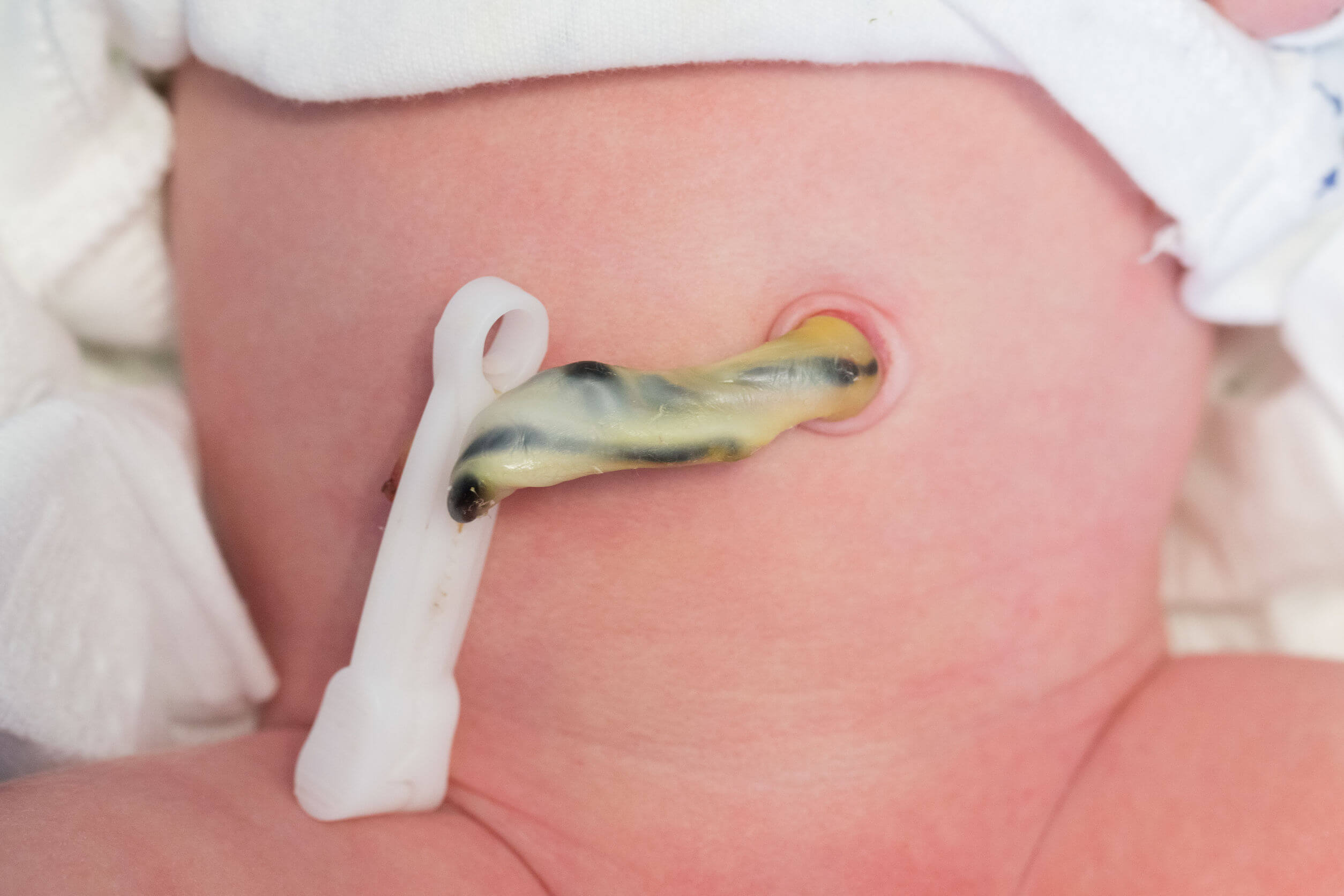
Usually, the stump (or remnant) of the umbilical cord dries up and falls off within 7 to 10 days after birth. However, if this process is prolonged and takes more than 15 days to come off, it is called a delayed umbilical cord separation.
In those babies without any underlying disease, this phenomenon may be related to some inadequate hygiene practices or to an alteration in the adhesion of white blood cells. The latter isn’t a problem in itself, however, the evaluation of the pediatrician is essential.
Other times, delayed umbilical cord separation is part of a range of clinical manifestations of a disease, such as congenital hypothyroidism.
2. Umbilical Hernia

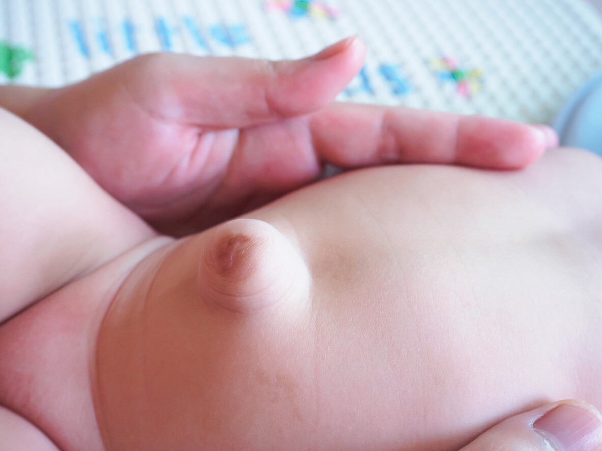
A naval hernia is an opening in the abdominal wall that arises as a result of a defect in the closure of the umbilical ring. This causes some abdominal organs and tissues to protrude out of the cavity, through the mentioned hole, and are only covered by the skin.
The size of hernias is variable, and in some babies, the bulge becomes visible on exertion (coughing, crying, or defecation). It’s usually detected between the second and third week of life after the stump falls off.
When the hernia is very large and covered only by a very thin, transparent layer of tissue, it’s known as an omphalocele.
It’s estimated that 8 out of 10 umbilical hernias close on their own before the age of 4. If this doesn’t occur, surgical closure may be necessary, depending on the case.
3. Omphalitis
Omphalitis is the infection of the umbilical cord and is a potentially serious health condition, which usually affects newborns in the first week of life. It usually has mild symptoms, but if it’s not treated in time, it can get worse. This occurs because the newborn doesn’t have the necessary defenses to deal with microbes.
The first cause of omphalitis is the colonization of bacteria in the stump of the cord, a product of poor hygiene during diaper changes or umbilical care. The most frequently implicated germs are Staphylococcus aureus (which lives on the skin) and Escherichia coli (which constitutes the intestinal flora).
Signs and symptoms of this condition include redness and swelling of the skin around the navel, pain and warmth to the touch, and passage of a foul-smelling, yellowish discharge through the cord.
The treatment of omphalitis is based on the use of antibiotics to eliminate the bacteria that produce it, in addition to measures to control symptoms, such as analgesics and fever reducers.
4. Umbilical Granuloma
An umbilical granuloma is another of the anomalies that can occur in the newborn’s belly button. This is formed as a result of excessive growth of scar tissue after the detachment of the umbilical cord.
It shows up as a small, reddish, shiny lump in the scar area. In general, it measures between 0.5 and 1 cm in diameter and doesn’t hurt.
To treat this condition, you can resort to the application of silver nitrate, a solution that favors the recovery of the affected tissue. Following this procedure, daily wound healing is performed with a 70% alcohol solution for one week.
5. Cutaneous navel and proboscis
This anomaly of the newborn belly button consists of a 1-3 cm flap of skin, which protrudes through the scar after the separation of the remaining cord.
When the segment is short, the defect shrinks over time and becomes part of the scar itself. This is known as a cutaneous navel. On the other hand, when the flap of skin is long and forms a kind of tube that doesn’t disappear, it’s called a proboscis navel and requires surgical correction.
6. Umbilical polyp
This condition is often confused with umbilical granuloma, although in this case, there’s a persistence of the omphalomesenteric duct within the defect. Because this structure must be completely closed after birth, an umbilical polyp usually requires surgery.

Check the condition of your newborn’s belly button every day
As you’ve seen, a newborn’s belly button can also get sick. For this reason, it’s important to assess it daily, at each diaper change, in order to detect any atypical trait.
At the same time, it’s essential to ensure adequate hygiene when bathing the child and when curing the umbilical cord to prevent complications.
In case of observing a delay in the healing of the umbilical cord, redness in the skin that surrounds it, or a lump in the navel scar, prompt evaluation by the pediatrician is recommended.
The umbilical cord is the structure that unites the baby with its mother during pregnancy and allows it to be nourished and oxygenated inside the uterus. At birth, this physical union is severed and later on, a scar will give way to the navel we all know. But did you know that some abnormalities in the newborn’s belly button can occur?
During the period of drying and falling off of the remaining stump of the cord, inconveniences can arise, some of which are potentially serious. For this reason, today we want to talk to you about abnormalities in a newborn’s belly button so that you recognize them and know how to act when you detect them.
1. Delayed detachment of the umbilical cord
Usually, the stump (or remnant) of the umbilical cord dries up and falls off within 7 to 10 days after birth. However, if this process is prolonged and takes more than 15 days to come off, it is called a delayed umbilical cord separation.
In those babies without any underlying disease, this phenomenon may be related to some inadequate hygiene practices or to an alteration in the adhesion of white blood cells. The latter isn’t a problem in itself, however, the evaluation of the pediatrician is essential.
Other times, delayed umbilical cord separation is part of a range of clinical manifestations of a disease, such as congenital hypothyroidism.
2. Umbilical Hernia

A naval hernia is an opening in the abdominal wall that arises as a result of a defect in the closure of the umbilical ring. This causes some abdominal organs and tissues to protrude out of the cavity, through the mentioned hole, and are only covered by the skin.
The size of hernias is variable, and in some babies, the bulge becomes visible on exertion (coughing, crying, or defecation). It’s usually detected between the second and third week of life after the stump falls off.
When the hernia is very large and covered only by a very thin, transparent layer of tissue, it’s known as an omphalocele.
It’s estimated that 8 out of 10 umbilical hernias close on their own before the age of 4. If this doesn’t occur, surgical closure may be necessary, depending on the case.
3. Omphalitis
Omphalitis is the infection of the umbilical cord and is a potentially serious health condition, which usually affects newborns in the first week of life. It usually has mild symptoms, but if it’s not treated in time, it can get worse. This occurs because the newborn doesn’t have the necessary defenses to deal with microbes.
The first cause of omphalitis is the colonization of bacteria in the stump of the cord, a product of poor hygiene during diaper changes or umbilical care. The most frequently implicated germs are Staphylococcus aureus (which lives on the skin) and Escherichia coli (which constitutes the intestinal flora).
Signs and symptoms of this condition include redness and swelling of the skin around the navel, pain and warmth to the touch, and passage of a foul-smelling, yellowish discharge through the cord.
The treatment of omphalitis is based on the use of antibiotics to eliminate the bacteria that produce it, in addition to measures to control symptoms, such as analgesics and fever reducers.
4. Umbilical Granuloma
An umbilical granuloma is another of the anomalies that can occur in the newborn’s belly button. This is formed as a result of excessive growth of scar tissue after the detachment of the umbilical cord.
It shows up as a small, reddish, shiny lump in the scar area. In general, it measures between 0.5 and 1 cm in diameter and doesn’t hurt.
To treat this condition, you can resort to the application of silver nitrate, a solution that favors the recovery of the affected tissue. Following this procedure, daily wound healing is performed with a 70% alcohol solution for one week.
5. Cutaneous navel and proboscis
This anomaly of the newborn belly button consists of a 1-3 cm flap of skin, which protrudes through the scar after the separation of the remaining cord.
When the segment is short, the defect shrinks over time and becomes part of the scar itself. This is known as a cutaneous navel. On the other hand, when the flap of skin is long and forms a kind of tube that doesn’t disappear, it’s called a proboscis navel and requires surgical correction.
6. Umbilical polyp
This condition is often confused with umbilical granuloma, although in this case, there’s a persistence of the omphalomesenteric duct within the defect. Because this structure must be completely closed after birth, an umbilical polyp usually requires surgery.

Check the condition of your newborn’s belly button every day
As you’ve seen, a newborn’s belly button can also get sick. For this reason, it’s important to assess it daily, at each diaper change, in order to detect any atypical trait.
At the same time, it’s essential to ensure adequate hygiene when bathing the child and when curing the umbilical cord to prevent complications.
In case of observing a delay in the healing of the umbilical cord, redness in the skin that surrounds it, or a lump in the navel scar, prompt evaluation by the pediatrician is recommended.
All cited sources were thoroughly reviewed by our team to ensure their quality, reliability, currency, and validity. The bibliography of this article was considered reliable and of academic or scientific accuracy.
- Araneda, L. y cols (2015). Patología del ombligo. Revista Pediatría Electrónica. Vol 12, N° 1. ISSN 0718-0918.
- Asociación española de pediatría (2008). Patología Umbilical Frecuente. Protocolos Diagnóstico Terapeúticos de la AEP: Neonatología. Recuperado de: https://www.aeped.es/sites/default/files/documentos/41.pdf
- García, A y cols (2019) Patología del área umbilical. Medicina clínica pediátrica. Vol 2, Nº 6, pp. 105-108.
- Novoa, A. (2004) El pediatra ante un lactante con caída tardía del cordón umbilical. Archivos argentinos de pediatría. Vol. 102 Nº Jul./jun. 2004. Disponible en: http://www.scielo.org.ar/scielo.php?script=sci_arttext&pid=S0325-00752004000300009#:~:text=Se%20considera%20ca%C3%ADda%20tard%C3%ADa%20del,de%20irrigaci%C3%B3n%20y%20la%20necrosis.
This text is provided for informational purposes only and does not replace consultation with a professional. If in doubt, consult your specialist.


