Anatomical and Physiological Changes During Labor


Written and verified by the pediatrician Marcela Alejandra Caffulli
Childbirth is the final stage of gestation, in which the woman gives birth to her baby and expels the placenta from the uterus. Regardless of the route through which the fetus leaves the maternal body, the female body undergoes very important anatomical and physiological changes during labor.
Just as at the beginning of pregnancy there were modifications to accommodate a new life, at this time, the phase of returning to the previous state begins. But not all structures will return to their original condition, as many of them will be put in place to ensure the care of the young.
Next, we’ll tell you how the body’s prepared in the moments before the arrival of the future baby.
The phases of labor
The baby’s birth is just one of the phases of labor. In fact, for this to occur, the maternal body needs to prepare in advance. Maternal, fetal, and hormonal factors participate in this process.
From the combination of the signals emitted by these three protagonists, the beginning of labor will result.
The phases of labor involve the entire set of anatomical and physiological changes in the woman’s body towards the end of gestation. In other words, it goes beyond the well-known stages of labor (dilation, expulsion, and delivery). For Cunningham et al. (2019), these phases include the following:
- Prelude to labor (or phase of inactivity): This involves much of the pregnancy and is characterized by remodeling of the cervix (without dilation).
- Preparation for labor (or uterine activation): Begins in the weeks before birth and includes the maturation of the cervix and some changes in the muscles of the uterus.
- Labor process (or uterine stimulation phase): This is the phase of painful uterine contractions, which are accompanied by the cardinal processes of labor (dilation, expulsion, and delivery).
- Recovery from childbirth (or involution): This begins after birth and involves the processes of uterine involution, repair of the cervix, and the beginning of lactation.

You may be interested in: Changes in Breasts During Pregnancy
Anatomical and physiological changes in the mother’s body during childbirth
As we’ve anticipated, the changes in the maternal organism follow a defined sequence to adapt to the needs of the new baby. As it matures, the female genital tract adapts to give birth and recover for the next pregnancy.
Next, we’ll describe the anatomical and physiological changes during labor that involve the main structures involved in the birth of the child.
Uterus and placenta
This is the cavity that houses the baby to ensure its nutrition, growth, and development until the mature fetal stage, around the 40th week of gestation.
During the 9 months, there are changes in all its structures: The endometrium (the uterine layer in contact with the fetal structures), the myometer (the muscular layer), and the cervix, among others.
Towards the end of pregnancy, the following changes occur:
- Cervical ripening: The cervix softens and shortens.
- Formation of the lower uterine segment: This region is made within the uterine cavity and is prepared to house the baby’s head. This way, it allows the fetus to fit into the maternal pelvis.
- Increased sensitivity of the myometrium to hormonal stimuli during labor (mainly oxytocin ) and an increased ability to contract.
When phase 3 (or the process of labor) begins, the uterine contractions necessary to expel the baby to the outside begin. These are usually intense, regular, and involuntary.
So, the rhythmic contraction of the myometrium causes the following changes:
- Effacement and cervical dilation (up to 10 centimeters in diameter)
- Loss of mucous plug
- Breaking of the amniotic sac
- Straightening of the spinal column of the fetus and descent through the birth canal
- Changes in the floor of the pelvis to favor the delivery of the baby
Once the baby leaves the uterus, this organ begins to shrink until it reaches the level of the navel (involution phase). This occurs thanks to the contraction of the myometrium but at levels al that are almost imperceptible for the mother. This mechanism seeks to compress the blood vessels in order to prevent bleeding.
At the same time, the placenta folds over itself and slowly detaches itself from the endometrium (or decidua). Within these two layers, a hematoma is formed with the remnants of blood and the entire organ is slowly released through the birth canal. This phenomenon is known as afterbirth.
Finally, the cervix enters a repair stage to avoid infection and to become fit again to house a new life.
Breasts
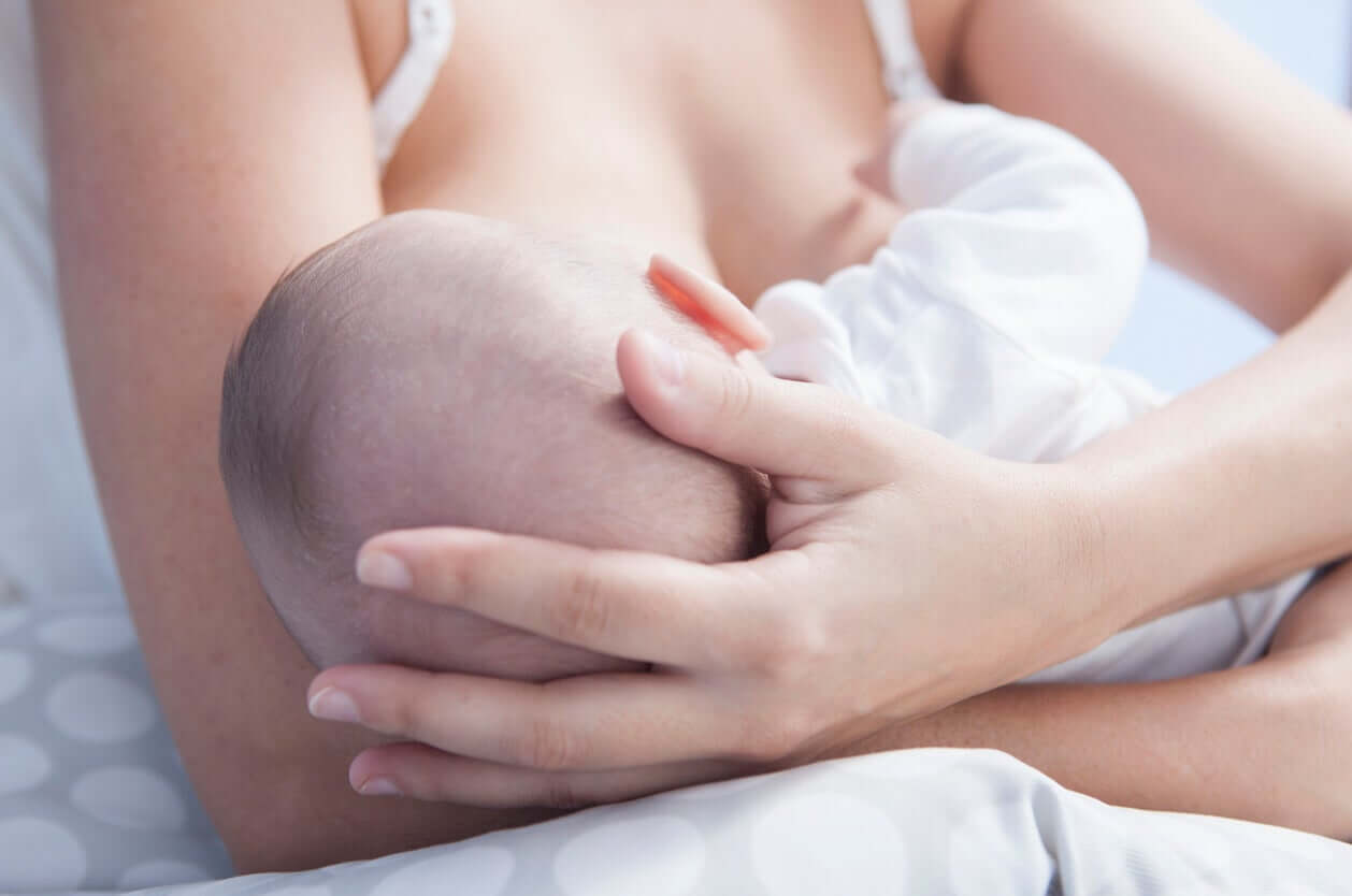
The new protagonists of this story will be the mammary glands, as they’ll have the important mission of nurturing the new baby.
Although their structures have developed during pregnancy, this machinery will start up after the baby’s first suckle. And just as with the genital organs, this whole process will depend on the corresponding hormonal stimuli.
Physical changes during labor are just the beginning…
Motherhood is much more than biology, and although these transformations are immense and palpable, they’re only the kickoff toward new life.
As you can see, nature is wise and shows us that bringing a child into the world isn’t an easy thing. On the contrary, it requires an effort in several aspects and a dedication of body and soul. But the prize that nature returns to us exceeds all expectations. Enjoy it!
Childbirth is the final stage of gestation, in which the woman gives birth to her baby and expels the placenta from the uterus. Regardless of the route through which the fetus leaves the maternal body, the female body undergoes very important anatomical and physiological changes during labor.
Just as at the beginning of pregnancy there were modifications to accommodate a new life, at this time, the phase of returning to the previous state begins. But not all structures will return to their original condition, as many of them will be put in place to ensure the care of the young.
Next, we’ll tell you how the body’s prepared in the moments before the arrival of the future baby.
The phases of labor
The baby’s birth is just one of the phases of labor. In fact, for this to occur, the maternal body needs to prepare in advance. Maternal, fetal, and hormonal factors participate in this process.
From the combination of the signals emitted by these three protagonists, the beginning of labor will result.
The phases of labor involve the entire set of anatomical and physiological changes in the woman’s body towards the end of gestation. In other words, it goes beyond the well-known stages of labor (dilation, expulsion, and delivery). For Cunningham et al. (2019), these phases include the following:
- Prelude to labor (or phase of inactivity): This involves much of the pregnancy and is characterized by remodeling of the cervix (without dilation).
- Preparation for labor (or uterine activation): Begins in the weeks before birth and includes the maturation of the cervix and some changes in the muscles of the uterus.
- Labor process (or uterine stimulation phase): This is the phase of painful uterine contractions, which are accompanied by the cardinal processes of labor (dilation, expulsion, and delivery).
- Recovery from childbirth (or involution): This begins after birth and involves the processes of uterine involution, repair of the cervix, and the beginning of lactation.

You may be interested in: Changes in Breasts During Pregnancy
Anatomical and physiological changes in the mother’s body during childbirth
As we’ve anticipated, the changes in the maternal organism follow a defined sequence to adapt to the needs of the new baby. As it matures, the female genital tract adapts to give birth and recover for the next pregnancy.
Next, we’ll describe the anatomical and physiological changes during labor that involve the main structures involved in the birth of the child.
Uterus and placenta
This is the cavity that houses the baby to ensure its nutrition, growth, and development until the mature fetal stage, around the 40th week of gestation.
During the 9 months, there are changes in all its structures: The endometrium (the uterine layer in contact with the fetal structures), the myometer (the muscular layer), and the cervix, among others.
Towards the end of pregnancy, the following changes occur:
- Cervical ripening: The cervix softens and shortens.
- Formation of the lower uterine segment: This region is made within the uterine cavity and is prepared to house the baby’s head. This way, it allows the fetus to fit into the maternal pelvis.
- Increased sensitivity of the myometrium to hormonal stimuli during labor (mainly oxytocin ) and an increased ability to contract.
When phase 3 (or the process of labor) begins, the uterine contractions necessary to expel the baby to the outside begin. These are usually intense, regular, and involuntary.
So, the rhythmic contraction of the myometrium causes the following changes:
- Effacement and cervical dilation (up to 10 centimeters in diameter)
- Loss of mucous plug
- Breaking of the amniotic sac
- Straightening of the spinal column of the fetus and descent through the birth canal
- Changes in the floor of the pelvis to favor the delivery of the baby
Once the baby leaves the uterus, this organ begins to shrink until it reaches the level of the navel (involution phase). This occurs thanks to the contraction of the myometrium but at levels al that are almost imperceptible for the mother. This mechanism seeks to compress the blood vessels in order to prevent bleeding.
At the same time, the placenta folds over itself and slowly detaches itself from the endometrium (or decidua). Within these two layers, a hematoma is formed with the remnants of blood and the entire organ is slowly released through the birth canal. This phenomenon is known as afterbirth.
Finally, the cervix enters a repair stage to avoid infection and to become fit again to house a new life.
Breasts

The new protagonists of this story will be the mammary glands, as they’ll have the important mission of nurturing the new baby.
Although their structures have developed during pregnancy, this machinery will start up after the baby’s first suckle. And just as with the genital organs, this whole process will depend on the corresponding hormonal stimuli.
Physical changes during labor are just the beginning…
Motherhood is much more than biology, and although these transformations are immense and palpable, they’re only the kickoff toward new life.
As you can see, nature is wise and shows us that bringing a child into the world isn’t an easy thing. On the contrary, it requires an effort in several aspects and a dedication of body and soul. But the prize that nature returns to us exceeds all expectations. Enjoy it!
All cited sources were thoroughly reviewed by our team to ensure their quality, reliability, currency, and validity. The bibliography of this article was considered reliable and of academic or scientific accuracy.
- Cunningham G, Leveno K, Bloom S et al. Fisiología del trabajo de parto. Capítulo 21. En: F. Gary Cunningham, Kenneth J. Leveno, Steven L. Bloom, Jodi S. Dashe, Barbara L. Hoffman, Brian M. Casey, Catherine Y. Spong. Williams Obstetricia. 25 edición. McGRAW-HILL INTERAMERICANA. 2019. ISBN: 978-1-4562-6736-0. Disponible en: https://accessmedicina.mhmedical.com/content.aspx?bookid=1525§ionid=100458658
- Condon JC, Jeyasuria P, Faust JM, Wilson JW, Mendelson CR. A decline in the levels of progesterone receptor coactivators in the pregnant uterus at term may antagonize progesterone receptor function and contribute to the initiation of parturition. Proc Natl Acad Sci U S A. 2003 Aug 5;100(16):9518-23. doi: 10.1073/pnas.1633616100. Epub 2003 Jul 28. PMID: 12886011; PMCID: PMC170950. Disponible en: https://www.ncbi.nlm.nih.gov/pmc/articles/PMC170950/
- Menon R, Bonney EA, Condon J, Mesiano S, Taylor RN. Novel concepts on pregnancy clocks and alarms: redundancy and synergy in human parturition. Hum Reprod Update. 2016 Sep;22(5):535-60. doi: 10.1093/humupd/dmw022. Epub 2016 Jun 30. PMID: 27363410; PMCID: PMC5001499. Disponible en: https://www.ncbi.nlm.nih.gov/pmc/articles/PMC5001499/
- Olson DM, Ammann C. Role of the prostaglandins in labour and prostaglandin receptor inhibitors in the prevention of preterm labour. Front Biosci. 2007 Jan 1;12:1329-43. doi: 10.2741/2151. PMID: 17127385. Disponible en: https://www.fbscience.com/Landmark/articles/10.2741/2151
This text is provided for informational purposes only and does not replace consultation with a professional. If in doubt, consult your specialist.


