10 Questions About Ultrasounds During Pregnancy


Written and verified by the nurse Leidy Mora Molina
During gestation, many doubts come to mind. Among them, there are questions related to ultrasounds during pregnancy. But what’s the frequency and function of these diagnostic tests? Learn more about them below!
Ultrasonography, also called ultrasound, is a diagnostic imaging method that uses high-frequency sound waves to observe internal tissues. In the case of the pregnant woman, it involves observing the growing baby.
The images obtained allow the specialist to see the baby’s anatomy and its development throughout the 9 months, as well as other structures such as the placenta and the amniotic fluid. In this way, possible complications can be diagnosed in a timely manner.
Discover the most frequent questions about ultrasounds during pregnancy and their answers
Ultrasounds during pregnancy are generally performed through the abdominal route. The exception is the first one, which is done before 10 weeks of gestation, in which the specialist uses the transvaginal route to carry it out.
However, as with everything in pregnancy, ultrasounds can generate some concerns in mothers. Below, we’ll address the most common doubts.
1. What’s the function of ultrasounds during pregnancy?
In the beginning, their function is to confirm the pregnancy, the location, the gestational age, the number of embryos, and the normality of the uterus and the tissues formed, such as the amniotic sac and the placenta.
Later, they allow for the evaluation of the baby’s growth, the fetal position, the location of the placenta, and the amount of amniotic fluid. For this reason, several ultrasounds are performed throughout gestation.
2. Which professional should perform ultrasounds during pregnancy?
This diagnostic procedure is only performed by professionals who are specialists in the area, such as obstetricians or radiologists. Observing and capturing these images requires professional training, so they’ll be performed by your own doctor or they’ll indicate a specific center where they can be performed.
3. Is ultrasound harmful to the mother and the fetus?
Another one of the most frequent questions about ultrasounds during pregnancy has to do with their safety. Ultrasounds are simple tests and aren’t harmful to the pregnancy. Because they work with ultrasound and don’t use ionizing radiation, such as X-rays, they’re considered safe during pregnancy.
Therefore, you shouldn’t be afraid to have ultrasound scans at this stage, because to date, no adverse effects caused by ultrasound have been detected either for the mother or for the baby.
4. What are the types of ultrasound?
Among the types of ultrasound scans performed during pregnancy are the following:
- 2D ultrasound: Images are seen in grayscale. This is the most commonly used by obstetricians.
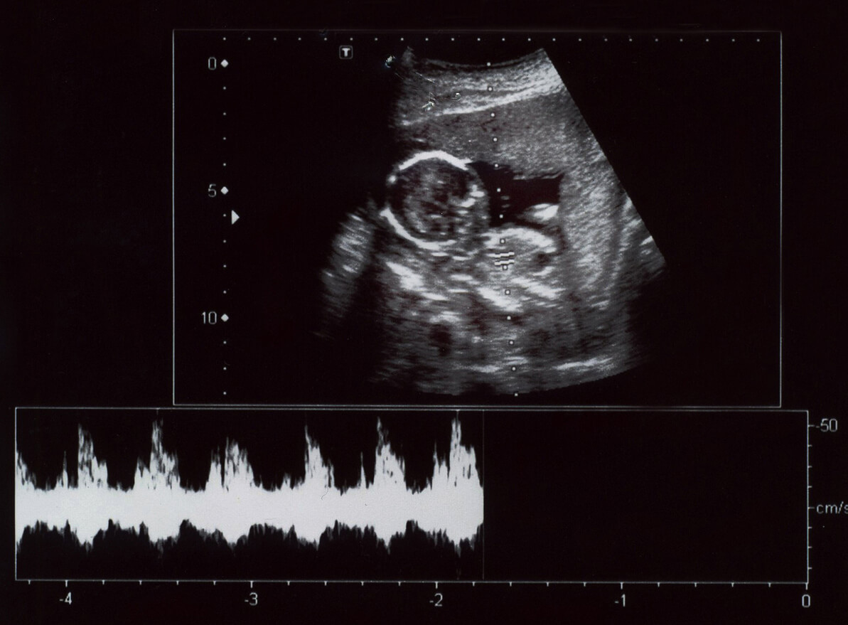
- Doppler ultrasound: Color Doppler is generally used for circulatory studies in veins and arteries, as fluids in movement can be visualized. Pulsed Doppler, on the other hand, studies blood vessels and the heart.
- 3D and 4D ultrasound: Through them, there’s better fetal visualization, and the shape and volume of the baby are clearly assessed. At the same time, the 4D ultrasound allows you to observe the baby’s movements in real time.
5. What prior care should I take before an ultrasound?
Because ultrasound is a simple procedure, you don’t need extensive prior preparation. However, it’s best not to apply moisturizing creams 24 hours before the test in the abdominal area, as some of them hinder the transmission of the ultrasound. It’s also important to wear comfortable clothing to make the process easier.
6. Should I have a full bladder for the ultrasound?
This requirement is important for the performance of any abdominal ultrasound in non-pregnant women. However, during pregnancy, it’s not necessary. This is because the baby’s surrounded by amniotic fluid that allows a good bounce of images to be visualized. In transvaginal ultrasounds, such as the one performed during the first trimester of pregnancy, having a full bladder isn’t necessary.
7. Can congenital alterations be observed in an ultrasound?
This depends on the genetic alteration that the fetus has. If it affects the anatomy of the affected organ or tissue, it’s observable, especially in 3D and 4D ultrasound scans. On the contrary, if the alteration only affects the function of an organ and doesn’t intervene in its morphology, then it can’t be detected by ultrasound.

8. How many ultrasound scans should I have during pregnancy?
The total number of ultrasound scans that are necessary during pregnancy depends on the evolution of the pregnancy. In this regard, the Spanish Society of Obstetrics and Gynecology recommends systematically performing 3 ultrasound scans in a low-risk pregnancy, distributed as follows:
- First ultrasound: Between 11 and 14 weeks, to detect embryonic vitality and the number of embryos
- Second ultrasound: Between 18 and 22 weeks, called morphological ultrasound
- Third ultrasound: Between 32 and 36 weeks, to observe the fetal position, the location of the placenta, and the amount of amniotic fluid
9. Are the results of ultrasounds reliable?
Although every day, technological advances allow for a better diagnostic range through ultrasound, some factors can affect the results. For example, the specialist’s experience, the fetal position, or the mother’s obesity, among others, can affect the correct interpretation. Even so, it’s considered a fairly reliable diagnostic test.
10. Should I be accompanied when I have an ultrasound?
This isn’t a procedure that requires accompaniment. However, mothers usually decide to go with their partner or another family member, so in most centers, an accompanying person is allowed.
The importance of ultrasound scans in pregnant women
Ultrasound scans throughout pregnancy serve as a guide for the physician to evaluate the evolution of the pregnancy. Also, in cases where treatable alterations are observed, the professional will be able to intervene in a timely manner to maintain the well-being of both mother and baby. In addition, they’ll be able to reassure the mother by knowing the baby’s condition.
In this regard, these studies are a very important diagnostic tool in prenatal care. They’re a useful and practical procedure that doesn’t put the health of the mother and child at risk. Just the same, if you still have questions about ultrasounds during pregnancy, be sure to discuss them with your doctor.
During gestation, many doubts come to mind. Among them, there are questions related to ultrasounds during pregnancy. But what’s the frequency and function of these diagnostic tests? Learn more about them below!
Ultrasonography, also called ultrasound, is a diagnostic imaging method that uses high-frequency sound waves to observe internal tissues. In the case of the pregnant woman, it involves observing the growing baby.
The images obtained allow the specialist to see the baby’s anatomy and its development throughout the 9 months, as well as other structures such as the placenta and the amniotic fluid. In this way, possible complications can be diagnosed in a timely manner.
Discover the most frequent questions about ultrasounds during pregnancy and their answers
Ultrasounds during pregnancy are generally performed through the abdominal route. The exception is the first one, which is done before 10 weeks of gestation, in which the specialist uses the transvaginal route to carry it out.
However, as with everything in pregnancy, ultrasounds can generate some concerns in mothers. Below, we’ll address the most common doubts.
1. What’s the function of ultrasounds during pregnancy?
In the beginning, their function is to confirm the pregnancy, the location, the gestational age, the number of embryos, and the normality of the uterus and the tissues formed, such as the amniotic sac and the placenta.
Later, they allow for the evaluation of the baby’s growth, the fetal position, the location of the placenta, and the amount of amniotic fluid. For this reason, several ultrasounds are performed throughout gestation.
2. Which professional should perform ultrasounds during pregnancy?
This diagnostic procedure is only performed by professionals who are specialists in the area, such as obstetricians or radiologists. Observing and capturing these images requires professional training, so they’ll be performed by your own doctor or they’ll indicate a specific center where they can be performed.
3. Is ultrasound harmful to the mother and the fetus?
Another one of the most frequent questions about ultrasounds during pregnancy has to do with their safety. Ultrasounds are simple tests and aren’t harmful to the pregnancy. Because they work with ultrasound and don’t use ionizing radiation, such as X-rays, they’re considered safe during pregnancy.
Therefore, you shouldn’t be afraid to have ultrasound scans at this stage, because to date, no adverse effects caused by ultrasound have been detected either for the mother or for the baby.
4. What are the types of ultrasound?
Among the types of ultrasound scans performed during pregnancy are the following:
- 2D ultrasound: Images are seen in grayscale. This is the most commonly used by obstetricians.
- Doppler ultrasound: Color Doppler is generally used for circulatory studies in veins and arteries, as fluids in movement can be visualized. Pulsed Doppler, on the other hand, studies blood vessels and the heart.
- 3D and 4D ultrasound: Through them, there’s better fetal visualization, and the shape and volume of the baby are clearly assessed. At the same time, the 4D ultrasound allows you to observe the baby’s movements in real time.
5. What prior care should I take before an ultrasound?
Because ultrasound is a simple procedure, you don’t need extensive prior preparation. However, it’s best not to apply moisturizing creams 24 hours before the test in the abdominal area, as some of them hinder the transmission of the ultrasound. It’s also important to wear comfortable clothing to make the process easier.
6. Should I have a full bladder for the ultrasound?
This requirement is important for the performance of any abdominal ultrasound in non-pregnant women. However, during pregnancy, it’s not necessary. This is because the baby’s surrounded by amniotic fluid that allows a good bounce of images to be visualized. In transvaginal ultrasounds, such as the one performed during the first trimester of pregnancy, having a full bladder isn’t necessary.
7. Can congenital alterations be observed in an ultrasound?
This depends on the genetic alteration that the fetus has. If it affects the anatomy of the affected organ or tissue, it’s observable, especially in 3D and 4D ultrasound scans. On the contrary, if the alteration only affects the function of an organ and doesn’t intervene in its morphology, then it can’t be detected by ultrasound.
8. How many ultrasound scans should I have during pregnancy?
The total number of ultrasound scans that are necessary during pregnancy depends on the evolution of the pregnancy. In this regard, the Spanish Society of Obstetrics and Gynecology recommends systematically performing 3 ultrasound scans in a low-risk pregnancy, distributed as follows:
- First ultrasound: Between 11 and 14 weeks, to detect embryonic vitality and the number of embryos
- Second ultrasound: Between 18 and 22 weeks, called morphological ultrasound
- Third ultrasound: Between 32 and 36 weeks, to observe the fetal position, the location of the placenta, and the amount of amniotic fluid
9. Are the results of ultrasounds reliable?
Although every day, technological advances allow for a better diagnostic range through ultrasound, some factors can affect the results. For example, the specialist’s experience, the fetal position, or the mother’s obesity, among others, can affect the correct interpretation. Even so, it’s considered a fairly reliable diagnostic test.
10. Should I be accompanied when I have an ultrasound?
This isn’t a procedure that requires accompaniment. However, mothers usually decide to go with their partner or another family member, so in most centers, an accompanying person is allowed.
The importance of ultrasound scans in pregnant women
Ultrasound scans throughout pregnancy serve as a guide for the physician to evaluate the evolution of the pregnancy. Also, in cases where treatable alterations are observed, the professional will be able to intervene in a timely manner to maintain the well-being of both mother and baby. In addition, they’ll be able to reassure the mother by knowing the baby’s condition.
In this regard, these studies are a very important diagnostic tool in prenatal care. They’re a useful and practical procedure that doesn’t put the health of the mother and child at risk. Just the same, if you still have questions about ultrasounds during pregnancy, be sure to discuss them with your doctor.
All cited sources were thoroughly reviewed by our team to ensure their quality, reliability, currency, and validity. The bibliography of this article was considered reliable and of academic or scientific accuracy.
- Gonzales, A (2009). Ecografía en obstetricia. España. Anales de Pediatría Continuada Vol. 7. Núm. 1. Páginas 39-44 (Febrero 2009).
- Nazario, C. (2011). La importancia de la ecografía a las 11+0 a 13+6 semanas de embarazo. Actualización. Perú. An Fac med. 2011;72(3):211-5.
- USAID (2018). Recomendaciones de la OMS sobre atención prenatal para una experiencia positiva del embarazo: Ecografía. Recuperado de: https://www.mcsprogram.org/wp-content/uploads/2018/07/WHO-MCSP-UltrasoundBriefer_A4_SP.pdf
This text is provided for informational purposes only and does not replace consultation with a professional. If in doubt, consult your specialist.


