What Is an Obstetric Ultrasound and What Is It Used For?


Written and verified by the nurse Leidy Mora Molina
An obstetric ultrasound is a technique of great importance for the control of pregnancies. Its clarity in showing the evolution of the baby and its practicality make it the key diagnostic test during this stage.
Performing ultrasounds during gestation allows the evaluation of fetal growth, in addition to the detection of possible malformations and problems in gestation. According to a study, it’s estimated that with this technique, it’s possible to diagnose between 65 and 75% of the malformations that complicate pregnancies.
This test is performed by an obstetrician-gynecologist or by an ultrasound specialist. It’s best to perform at least 3 ultrasound scans during pregnancy. Keep reading to learn more about this diagnostic method.
What’s an obstetric ultrasound and how does it work?
An obstetric ultrasound or obstetric ultrasound is a diagnostic test where, through images, the evaluation of the fetus and its surrounding structures is achieved. Ultrasound is used for this, that is, high-frequency sound waves to reproduce images of our interior, in this case, of the developing baby and the surrounding tissues.
The procedure is performed through a device called a transducer, which is responsible for sending ultrasound waves that hit the tissues to be observed. It picks up the waves sent from the inside and converts them into images.
The evolution of this device has allowed images to be sharper and even include movement. There are different modalities for this:
- 2D ultrasound: Images are observed in 2 dimensions and in grayscale. It’s the most commonly used.
- Doppler ultrasound: Studies the blood vessels and the heart. In pulsed Doppler, the pattern of blood flow in a specific area is observed.
- 3D ultrasound: Allows the volume to be seen, which is ideal for a better fetal evaluation.
- Echosonogram or 4D ultrasound: Volume and movement are seen live.
- 5D ultrasound: The same as in the 4D, but in color and with greater clarity.
Types of obstetric ultrasound
Depending on the size of the uterus and the tissue to be studied, obstetric ultrasounds can be performed in two ways:
- Transvaginal ultrasound: The imaging transducer is introduced through the vagina in order to evaluate structures close to the vagina. It’s used to evaluate pregnancy in the first trimester because the embryo’s still very small. It’s also used to evaluate and measure other structures such as the cervix.
- Abdominal ultrasound: The convex transducer is placed on the skin of the abdomen. It’s used from the second trimester onwards.
What’s an obstetric ultrasound for?
An obstetric ultrasound is used to assess certain important points in pregnancy and its evolution throughout the 40 weeks, among which the following stand out:
- Fetal viability and heart rate of the baby
- Determining the number of fetuses and their chorionicity
- Gestational age
- Identifying problems related to the placenta, uterus, cervix, and ovaries
- Observing the baby’s anatomy and growth during gestation
- Fetal position within the uterus
- The evaluation of possible chromosomal abnormalities
When should they be performed?
The Spanish Society of Obstetrics and Gynecology recommends performing 3 ultrasounds during gestation, that is, one in each trimester of pregnancy. The first one is done between 10 and 14 weeks of gestation; the second, between 18 and 22 weeks; and the third, between 34 and 36 weeks.

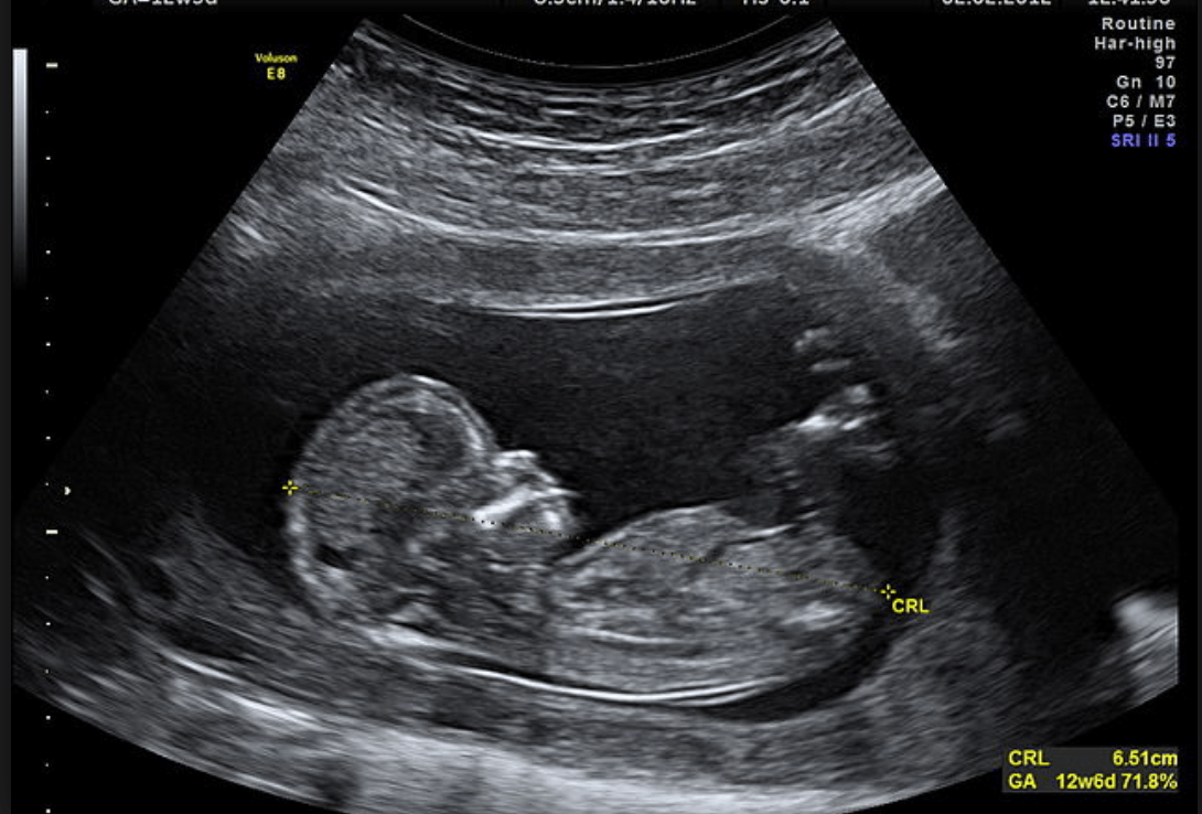
First-trimester ultrasound
This ultrasound is performed in order to observe if the gestation is viable. It observes if there’s a live embryo, if there’s an amniotic sac, and the number of embryos present. Also, the size of the fetus is measured, from the skull to the coccyx, to determine the time of gestation.
Second-trimester ultrasound
In the second trimester diagnosis, the fetal morphology is evaluated, that is, the evaluation of all the baby’s organs, such as the structures of the brain, heart, and abdominal organs. Other structures such as the placenta are also evaluated.
Third-trimester ultrasound
In this case, the position of the fetus, its viability, and growth are analyzed. Also, the insertion of the placenta and the amount of amniotic fluid surrounding the baby.
Can ultrasound affect the baby’s health?
Obstetric ultrasounds are a non-invasive and very safe technique to be performed during gestation. They have no risk, as they don’t use ionizing radiation. They also have no known harmful effects on the mother or the baby. This test doesn’t require prior preparation and usually lasts between 15 and 20 minutes, making it practical and safe for assessing the well-being of the pregnancy.
Physicians may indicate more frequent ultrasound scans, especially if the mother is anxious to see the evolution of the baby or if a problem has been detected in the pregnancy.
Obstetric ultrasound is a technological advance that allows the specialist to make timely therapeutic decisions. It also represents a beautiful moment for the mother, as she can observe the baby growing in her womb and foster bonding from before birth.
An obstetric ultrasound is a technique of great importance for the control of pregnancies. Its clarity in showing the evolution of the baby and its practicality make it the key diagnostic test during this stage.
Performing ultrasounds during gestation allows the evaluation of fetal growth, in addition to the detection of possible malformations and problems in gestation. According to a study, it’s estimated that with this technique, it’s possible to diagnose between 65 and 75% of the malformations that complicate pregnancies.
This test is performed by an obstetrician-gynecologist or by an ultrasound specialist. It’s best to perform at least 3 ultrasound scans during pregnancy. Keep reading to learn more about this diagnostic method.
What’s an obstetric ultrasound and how does it work?
An obstetric ultrasound or obstetric ultrasound is a diagnostic test where, through images, the evaluation of the fetus and its surrounding structures is achieved. Ultrasound is used for this, that is, high-frequency sound waves to reproduce images of our interior, in this case, of the developing baby and the surrounding tissues.
The procedure is performed through a device called a transducer, which is responsible for sending ultrasound waves that hit the tissues to be observed. It picks up the waves sent from the inside and converts them into images.
The evolution of this device has allowed images to be sharper and even include movement. There are different modalities for this:
- 2D ultrasound: Images are observed in 2 dimensions and in grayscale. It’s the most commonly used.
- Doppler ultrasound: Studies the blood vessels and the heart. In pulsed Doppler, the pattern of blood flow in a specific area is observed.
- 3D ultrasound: Allows the volume to be seen, which is ideal for a better fetal evaluation.
- Echosonogram or 4D ultrasound: Volume and movement are seen live.
- 5D ultrasound: The same as in the 4D, but in color and with greater clarity.
Types of obstetric ultrasound
Depending on the size of the uterus and the tissue to be studied, obstetric ultrasounds can be performed in two ways:
- Transvaginal ultrasound: The imaging transducer is introduced through the vagina in order to evaluate structures close to the vagina. It’s used to evaluate pregnancy in the first trimester because the embryo’s still very small. It’s also used to evaluate and measure other structures such as the cervix.
- Abdominal ultrasound: The convex transducer is placed on the skin of the abdomen. It’s used from the second trimester onwards.
What’s an obstetric ultrasound for?
An obstetric ultrasound is used to assess certain important points in pregnancy and its evolution throughout the 40 weeks, among which the following stand out:
- Fetal viability and heart rate of the baby
- Determining the number of fetuses and their chorionicity
- Gestational age
- Identifying problems related to the placenta, uterus, cervix, and ovaries
- Observing the baby’s anatomy and growth during gestation
- Fetal position within the uterus
- The evaluation of possible chromosomal abnormalities
When should they be performed?
The Spanish Society of Obstetrics and Gynecology recommends performing 3 ultrasounds during gestation, that is, one in each trimester of pregnancy. The first one is done between 10 and 14 weeks of gestation; the second, between 18 and 22 weeks; and the third, between 34 and 36 weeks.
First-trimester ultrasound
This ultrasound is performed in order to observe if the gestation is viable. It observes if there’s a live embryo, if there’s an amniotic sac, and the number of embryos present. Also, the size of the fetus is measured, from the skull to the coccyx, to determine the time of gestation.
Second-trimester ultrasound
In the second trimester diagnosis, the fetal morphology is evaluated, that is, the evaluation of all the baby’s organs, such as the structures of the brain, heart, and abdominal organs. Other structures such as the placenta are also evaluated.
Third-trimester ultrasound
In this case, the position of the fetus, its viability, and growth are analyzed. Also, the insertion of the placenta and the amount of amniotic fluid surrounding the baby.
Can ultrasound affect the baby’s health?
Obstetric ultrasounds are a non-invasive and very safe technique to be performed during gestation. They have no risk, as they don’t use ionizing radiation. They also have no known harmful effects on the mother or the baby. This test doesn’t require prior preparation and usually lasts between 15 and 20 minutes, making it practical and safe for assessing the well-being of the pregnancy.
Physicians may indicate more frequent ultrasound scans, especially if the mother is anxious to see the evolution of the baby or if a problem has been detected in the pregnancy.
Obstetric ultrasound is a technological advance that allows the specialist to make timely therapeutic decisions. It also represents a beautiful moment for the mother, as she can observe the baby growing in her womb and foster bonding from before birth.
All cited sources were thoroughly reviewed by our team to ensure their quality, reliability, currency, and validity. The bibliography of this article was considered reliable and of academic or scientific accuracy.
- Gonzales, A. (2009). Ecografía en obstetricia. Anales de pediatría continuada. Vol. 7. Núm. 1. páginas 39-44 (Febrero 2009)
- Lebit F. (2011). The Role of 4D Ultrasound in the Assessment of Fetal Behaviour. Maedica a journal of clinical medicine. 2011 Apr; 6(2): 120–127.
- Sociedad Argentina de Ultrasonografía en medicina y biología (2016) Guía para el estudio morfológico del segundo trimestre del embarazo. Argentina. Revista Argentina de Ultrasonido – 2016; Volumen 15 N°1: 372 – 379.
- Sociedad Española de ginecología y obstetricia (2018). Control prenatal del embarazo normal. Prog Obstet Ginecol 2018;61(5):510-527.
This text is provided for informational purposes only and does not replace consultation with a professional. If in doubt, consult your specialist.


