What's The Importance of Morphological Ultrasound


Written and verified by the pediatrician Marcela Alejandra Caffulli
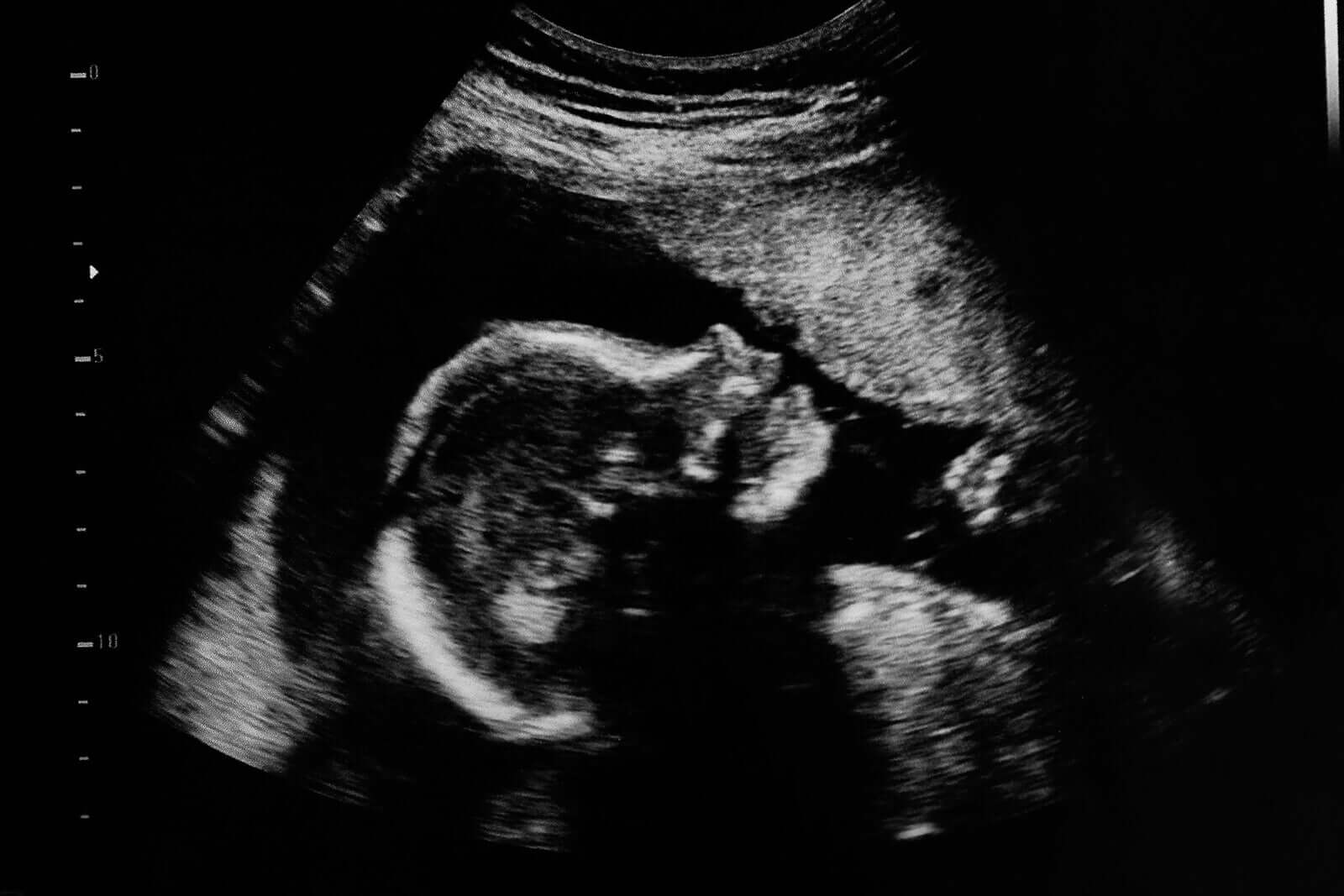
What is a morphological ultrasound?
A morphological ultrasound is a non-invasive study that seeks to evaluate how the baby’s body develops inside the mother’s womb. Through it, it’s possible to detect certain fetal pathologies such as congenital malformations in a timely manner.
It involves the use of a special device that emits high-frequency sound waves into the woman’s abdomen.
The sound disperses through the amniotic fluid and “collides” with the different structures of the baby. This generates a response signal (echo), which a computer captures and displays as a 2- or 3-dimensional image of the baby.
Ultrasounds serve many purposes throughout the pregnancy. However, the main objective of second-trimester ultrasounds is to evaluate how the different organs of the baby have formed.
Next, we’ll explain in detail the purposes of morphological ultrasound (Puerto, 2018):
- Evaluating the baby’s anatomy: It confirms that the formation of the organs is correct and that the fetal structures maintain the correct proportions. If the specialist detects any malformations, they’ll complete evaluation with more specialized studies.
- Observing fetal growth: Depending on the gestational age of the baby, it determines if the growth rate is appropriate or if the baby has intrauterine growth retardation.
- Calculating the volume of amniotic fluid: By means of some measurement parameters, such as the amniotic fluid index (AMI), it’s possible to detect the presence of an excess (polyhydramnios) or deficit (oligohydramnios) of amniotic fluid.
- Examining the condition of the placenta: Mainly, the location of this organ with respect to the internal cervical orifice and the site where the umbilical cord is inserted.
When to perform a morphological ultrasound
By the middle of the second trimester of pregnancy, most of the organs and structures of the fetus are already sufficiently formed. For this reason, it’s considered the optimal time to perform the morphological ultrasound.
Specialists recommend carrying out this study between week 18 and week 20 of gestation. In this period of time, three factors coincide that determine the success of the study:
- The baby’s bones still don’t have much calcium and this allows the correct examination of the fetal internal structures.
- The amount of amniotic fluid is adequate so that the sound waves distribute around the entire surface of the fetus.
- The size of the baby is large enough to observe in detail how its organs have formed.
Facts you should know
Morphological ultrasound is useful in the detection of different malformations or congenital anomalies in the fetus in time. Although not all these diseases are known, their frequency of appearance is not negligible.
According to estimates, 1 in 33 children born in the world suffers from a congenital defect and more than 3 million children suffer from a disability due to this cause (PAHO, 2015).
Although ultrasound can’t prevent the appearance of any of these conditions, it offers families the possibility of receiving the necessary treatments to reduce the risk of death or complications in the health of babies.
For this reason, it’s extremely important to carry out this and other health checks during pregnancy, especially in high-risk pregnancies.
Does it hurt?
Without a doubt, the morphological ultrasound doesn’t cause pain for the mother or for her baby.
It takes place using ultrasound equipment, which consists of a computer and a signal transducer. The latter is what the ultrasound technician places on the skin of the mother’s belly.

To move the device from one side of the abdomen to the other, the professional uses a gel, which prevents any pain, itching, or unpleasant sensation.
At the same time, the sound waves that the machine emits into the mother’s uterus don’t cause pain or any suffering to the baby.
Does it involve any risks?
Ultrasounds are safe studies for both the mother and her child, as they don’t involve any present or future risk.
The pressures on the womb are mild and don’t increase the likelihood of complications from external trauma.
As for sound waves, they don’t pose a risk to the health of the fetus, as do x-rays.
Is it useful for making accurate diagnoses?
This is a reliable enough study, as long as it takes place using the appropriate equipment and the operator (usually a physician) has sufficient training.
For this reason, it’s said to be an “operator dependent” study, as its success depends on the human hand that carries it out.
Morphological ultrasound is a routine procedure that seeks to detect those pregnancies with a possible risk of congenital malformations.
If the specialist detects any alteration in the baby’s body, they’ll need to repeat the ultrasound after a few weeks. At the same time, they’ll indicate another type of more specific ultrasound. For example, those studies that include a fetal doppler.
Regarding the importance of morphological ultrasound
As we’ve emphasized throughout the article, ultrasounds are simple, non-invasive, and very safe studies for the mother and her baby.
From them, it’s possible to obtain relevant information at an opportune moment, in order to offer families the necessary advice and interventions, thus minimizing future risks.
Don’t forget that complying with all obstetric controls and routine studies is the best way to give good health to your unborn child.
What is a morphological ultrasound?
A morphological ultrasound is a non-invasive study that seeks to evaluate how the baby’s body develops inside the mother’s womb. Through it, it’s possible to detect certain fetal pathologies such as congenital malformations in a timely manner.
It involves the use of a special device that emits high-frequency sound waves into the woman’s abdomen.
The sound disperses through the amniotic fluid and “collides” with the different structures of the baby. This generates a response signal (echo), which a computer captures and displays as a 2- or 3-dimensional image of the baby.
Ultrasounds serve many purposes throughout the pregnancy. However, the main objective of second-trimester ultrasounds is to evaluate how the different organs of the baby have formed.
Next, we’ll explain in detail the purposes of morphological ultrasound (Puerto, 2018):
- Evaluating the baby’s anatomy: It confirms that the formation of the organs is correct and that the fetal structures maintain the correct proportions. If the specialist detects any malformations, they’ll complete evaluation with more specialized studies.
- Observing fetal growth: Depending on the gestational age of the baby, it determines if the growth rate is appropriate or if the baby has intrauterine growth retardation.
- Calculating the volume of amniotic fluid: By means of some measurement parameters, such as the amniotic fluid index (AMI), it’s possible to detect the presence of an excess (polyhydramnios) or deficit (oligohydramnios) of amniotic fluid.
- Examining the condition of the placenta: Mainly, the location of this organ with respect to the internal cervical orifice and the site where the umbilical cord is inserted.
When to perform a morphological ultrasound
By the middle of the second trimester of pregnancy, most of the organs and structures of the fetus are already sufficiently formed. For this reason, it’s considered the optimal time to perform the morphological ultrasound.
Specialists recommend carrying out this study between week 18 and week 20 of gestation. In this period of time, three factors coincide that determine the success of the study:
- The baby’s bones still don’t have much calcium and this allows the correct examination of the fetal internal structures.
- The amount of amniotic fluid is adequate so that the sound waves distribute around the entire surface of the fetus.
- The size of the baby is large enough to observe in detail how its organs have formed.
Facts you should know
Morphological ultrasound is useful in the detection of different malformations or congenital anomalies in the fetus in time. Although not all these diseases are known, their frequency of appearance is not negligible.
According to estimates, 1 in 33 children born in the world suffers from a congenital defect and more than 3 million children suffer from a disability due to this cause (PAHO, 2015).
Although ultrasound can’t prevent the appearance of any of these conditions, it offers families the possibility of receiving the necessary treatments to reduce the risk of death or complications in the health of babies.
For this reason, it’s extremely important to carry out this and other health checks during pregnancy, especially in high-risk pregnancies.
Does it hurt?
Without a doubt, the morphological ultrasound doesn’t cause pain for the mother or for her baby.
It takes place using ultrasound equipment, which consists of a computer and a signal transducer. The latter is what the ultrasound technician places on the skin of the mother’s belly.
To move the device from one side of the abdomen to the other, the professional uses a gel, which prevents any pain, itching, or unpleasant sensation.
At the same time, the sound waves that the machine emits into the mother’s uterus don’t cause pain or any suffering to the baby.
Does it involve any risks?
Ultrasounds are safe studies for both the mother and her child, as they don’t involve any present or future risk.
The pressures on the womb are mild and don’t increase the likelihood of complications from external trauma.
As for sound waves, they don’t pose a risk to the health of the fetus, as do x-rays.
Is it useful for making accurate diagnoses?
This is a reliable enough study, as long as it takes place using the appropriate equipment and the operator (usually a physician) has sufficient training.
For this reason, it’s said to be an “operator dependent” study, as its success depends on the human hand that carries it out.
Morphological ultrasound is a routine procedure that seeks to detect those pregnancies with a possible risk of congenital malformations.
If the specialist detects any alteration in the baby’s body, they’ll need to repeat the ultrasound after a few weeks. At the same time, they’ll indicate another type of more specific ultrasound. For example, those studies that include a fetal doppler.
Regarding the importance of morphological ultrasound
As we’ve emphasized throughout the article, ultrasounds are simple, non-invasive, and very safe studies for the mother and her baby.
From them, it’s possible to obtain relevant information at an opportune moment, in order to offer families the necessary advice and interventions, thus minimizing future risks.
Don’t forget that complying with all obstetric controls and routine studies is the best way to give good health to your unborn child.
All cited sources were thoroughly reviewed by our team to ensure their quality, reliability, currency, and validity. The bibliography of this article was considered reliable and of academic or scientific accuracy.
- Puerto B. Segundo trimestre. ecografía morfológica y cribado de patología. Capítulo 11. En: Gratacós E, Figueras F, Martínez JM. Medicina Fetal. 2da Edición. Barcelona: Panamericana. Año 2018.
- Richards D. Ecografía obstétrica: estudios de imagen, establecimiento de fechas, crecimiento y anomalías. Capítulo 9. En: Gabe S et al. Obstetricia. Embarazos normales y de riesgo. 7ma edición. España: Elsevier. Año 2019.
- Callen P, Norton M. Ecografía Obstétrica. Capítulo 1. En: Norton M. Callen. Ecografía en obstetricia y ginecología. 6ta edición. España: Elsevier. Año 2018.
- Reddy UM, Abuhamad AZ, Levine D. Saade GR. Fetal imaging: executive summary of a joint Eunice Kennedy Shriver National Institute of Child Health and Human Development, Society for Maternal-Fetal Medicine, American Institute of Ultrasound in Medicine, American College of Obstetricians and Gynecologists, American College of Radiology, Society for Pediatric Radiology, and Society of Radiologists in Ultrasound Fetal Imaging Workshop. Obstet Gynecol. 2014;123:1070-1082. Disponible en: https://pubmed.ncbi.nlm.nih.gov/24764329/
- American Academy of Pediatrics. Malformaciones congénitas. Genetics in Primary Care Institute – AAP. Healthy children. [Internet] Última actualización 2019. Disponible en: https://www.healthychildren.org/Spanish/health-issues/conditions/developmental-disabilities/Paginas/Congenital-Abnormalities.aspx
- Organización Panamericana de la Salud. Las anomalías congénitas son la segunda causa de muerte en los niños menores de 5 años en las Américas. [Internet] Marzo 2015. Disponible en: https://www3.paho.org/hq/index.php?option=com_content&view=article&id=10487:2015-anomalias-congenitas-segunda-causa-muerte-ninos-menores-5-anos-americas&Itemid=1926&lang=es
This text is provided for informational purposes only and does not replace consultation with a professional. If in doubt, consult your specialist.


