How Many Ultrasounds Are Necessary in Pregnancy?


Written and verified by the pediatrician Marcela Alejandra Caffulli
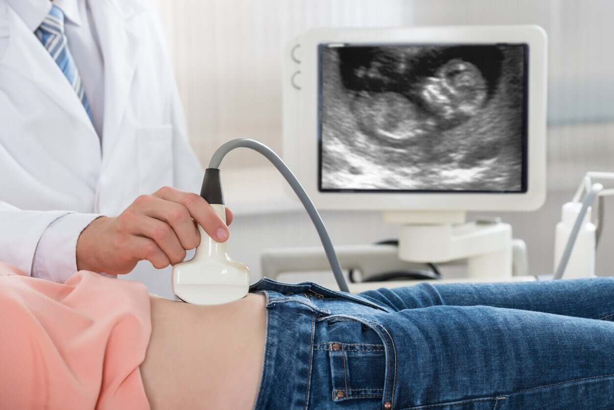
Current obstetric practice is far from that of just 50 years ago. The fact of the matter is that the implementation of ultrasounds as part of the routine of pregnancy controls has marked a before and after. But can there be too much of a good thing? How many ultrasounds are necessary during pregnancy?
Today, we have modern ultrasound machines and well-trained professionals to evaluate in detail the development of the fetus and diagnose (or treat) diseases in the early stages of pregnancy.
However, the dilemma that we’re presented with is the possibility of overusing this technique and adding unnecessary risk to the health of the baby or that of the mother. Therefore, today, we’re going to tell you how many ultrasounds are really necessary during your pregnancy and for what purpose they should be performed. Keep reading to learn everything you need to know.
Ultrasound scans in pregnancy: Benefits versus risks
All parents are excited to see their baby “swimming” inside the womb. Even more so, when the specialist conducting the study informs them that everything looks as expected. But have you ever wondered what would happen if the report was the opposite?
As with any complementary study, pregnancy ultrasounds give us valuable information about the health status of the mother and the baby. But this doesn’t mean that it’s the only form of evaluation or that it’s infallible. In this sense, it’s essential that those who practice it have sufficient training to interpret data and contextualize it appropriately.
Ultrasound technology has numerous advantages from its technical aspect: It doesn’t require excessively sophisticated equipment, it doesn’t emit radiation, it can be practiced throughout the entire pregnancy, and it’s safe for the fetus and its mother.
Among its disadvantages is the fact that it’s an “operator-dependent” study, which implies that the images (and data) obtained from the study depend on the human hand. Therefore, it’s important to know what information you want to know, how to look for it, how to interpret it, and above all, how to corroborate each finding that goes outside the normal parameters.
So, to get the most out of it, doctors need to make a rational and conscious use of ultrasounds. Likewise, we should avoid using them for recreational or entertainment purposes.
Necessary pregnancy ultrasounds
As we’ve mentioned, ultrasounds are safe to perform from the beginning of pregnancy, but they should be reserved for key moments and to evaluate specific issues.
International scientific societies have developed guidelines and consensus to establish the standards of safe practice. In accordance with this, the Spanish Society of Obstetrics and Gynecology (SEOG) recommends performing 3 ultrasound examinations in normal pregnancies, one per trimester.
First trimester
The SEOG recommends performing the first ultrasound between weeks 10 and 14 of pregnancy. This study is part of the combined screening of the first trimester and its main objective is to corroborate the presence of the embryo or fetus within the maternal uterus.
In addition, this ultrasound evaluates the number of gestational sacs (and babies), the location of the placenta, fetal cardiac activity, and measurements are made to know the gestational age with greater precision.
At this stage of pregnancy, it’s also possible to screen for malformations and some chromosome diseases, such as Down syndrome. Some of the ultrasound procedures used are nuchal translucency (TN) and nasal bone measurement, among others.
You may be interested in: Prenatal Tests: Safe, Fast, and Reliable
Second trimester
Between weeks 19 and 21, experts recommend performing the second ultrasound, which has the main objective of evaluating how all the structures of the baby have been formed. This type of study is known as a morphological ultrasound or fetal scan.
Thanks to the availability of high-resolution imaging equipment, it’s possible to carefully evaluate the different organs and systems of the fetus in order to detect early malformations that must be corrected in utero or shortly after birth.
In addition, in the case of diagnosing a complex genetic syndrome or any health condition that’s incompatible with life, the most appropriate conduct for each case may be taken.
Third trimester
The usefulness of ultrasound in the third trimester of pregnancy is controversial. Many scientific societies claim that the performance doesn’t offer any benefit to the health of the mother or the baby when it comes to low-risk pregnancies.
In fact, a recent Cochrane Collaboration systematic review showed that there are no clear benefits for women who have an ultrasound this trimester compared to those who don’t.
However, the SEGO recommends conducting this study in all pregnant women between weeks 32 and 36 (inclusive) with the following purpose:
- Checking the health status of the fetus: Position, vitality, and growth.
- Detecting abnormalities related to the position of the placenta in the uterus.
- Quantifying the volume of amniotic fluid to detect excess amounts or deficiencies.
- Assessing the blood supply to the placenta and blood flow through the cord when necessary.
Other pregnancy ultrasounds
In the case of high-risk pregnancies or when you want to observe a specific aspect in detail, you can use another type of ultrasound: Specialized or limited.
Regarding 3D or 4D ultrasounds, the suitability of their use in routine pregnancies is still a controversial issue.
Remember that sometimes “less is more”
Now that you know when and why your obstetrician will tell you to perform the different pregnancy ultrasounds, you can rest easy with the idea that 3 are enough.
Sometimes, doctors request more studies in one woman than in another, due to the particular health conditions of each one. In fact, it’s to be expected that you yourself will have different experiences in each of your own pregnancies, as each one will be unique.
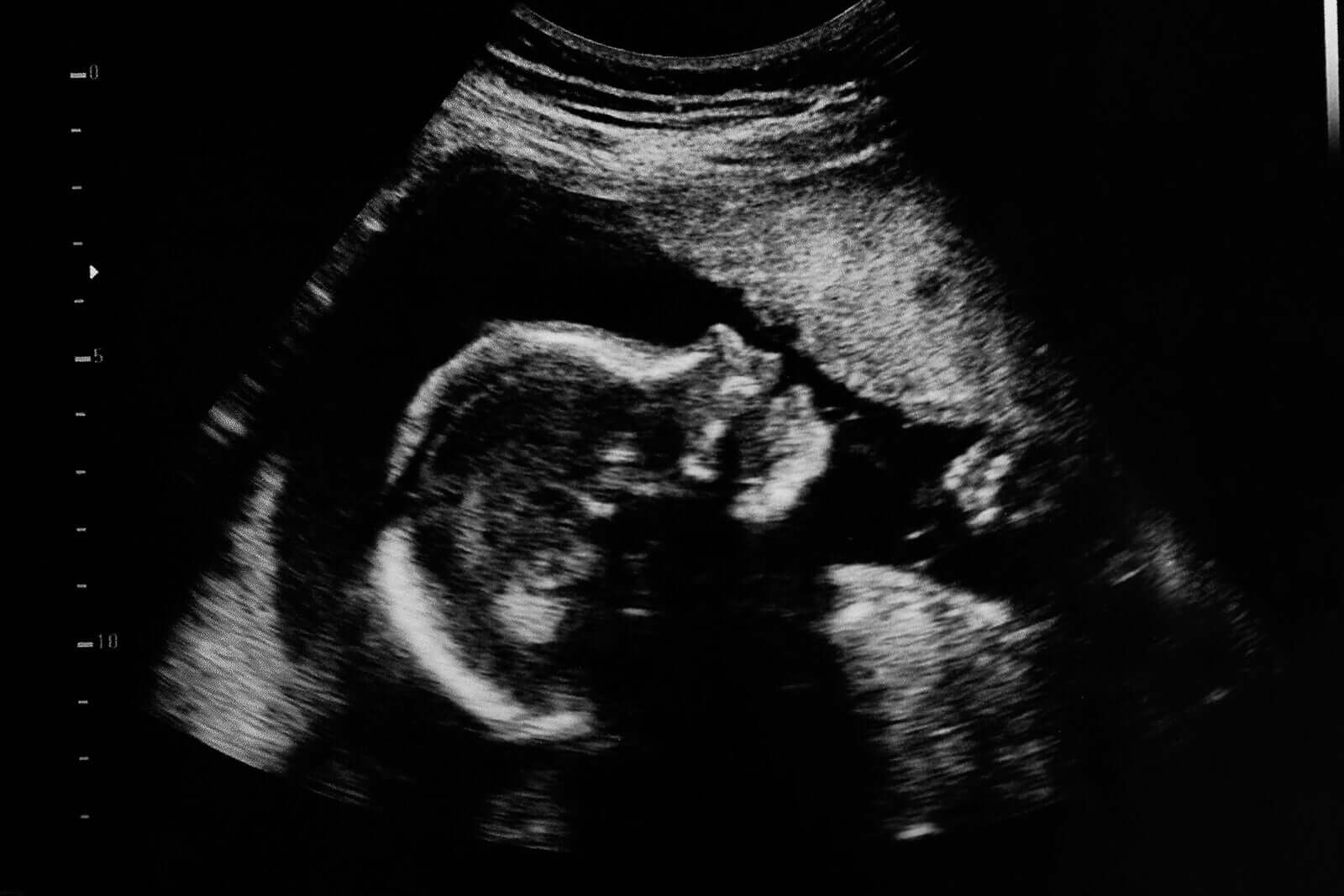
Beyond the beautiful detail of the photo in the foreground, you should always keep in mind that pregnancy ultrasounds are medical studies to diagnose precise issues. They’re not and shouldn’t be used in any way as photographic sessions of the unborn baby.
Remember that “less is more” and, in this case, this phrase applies better than ever.
Current obstetric practice is far from that of just 50 years ago. The fact of the matter is that the implementation of ultrasounds as part of the routine of pregnancy controls has marked a before and after. But can there be too much of a good thing? How many ultrasounds are necessary during pregnancy?
Today, we have modern ultrasound machines and well-trained professionals to evaluate in detail the development of the fetus and diagnose (or treat) diseases in the early stages of pregnancy.
However, the dilemma that we’re presented with is the possibility of overusing this technique and adding unnecessary risk to the health of the baby or that of the mother. Therefore, today, we’re going to tell you how many ultrasounds are really necessary during your pregnancy and for what purpose they should be performed. Keep reading to learn everything you need to know.
Ultrasound scans in pregnancy: Benefits versus risks
All parents are excited to see their baby “swimming” inside the womb. Even more so, when the specialist conducting the study informs them that everything looks as expected. But have you ever wondered what would happen if the report was the opposite?
As with any complementary study, pregnancy ultrasounds give us valuable information about the health status of the mother and the baby. But this doesn’t mean that it’s the only form of evaluation or that it’s infallible. In this sense, it’s essential that those who practice it have sufficient training to interpret data and contextualize it appropriately.
Ultrasound technology has numerous advantages from its technical aspect: It doesn’t require excessively sophisticated equipment, it doesn’t emit radiation, it can be practiced throughout the entire pregnancy, and it’s safe for the fetus and its mother.
Among its disadvantages is the fact that it’s an “operator-dependent” study, which implies that the images (and data) obtained from the study depend on the human hand. Therefore, it’s important to know what information you want to know, how to look for it, how to interpret it, and above all, how to corroborate each finding that goes outside the normal parameters.
So, to get the most out of it, doctors need to make a rational and conscious use of ultrasounds. Likewise, we should avoid using them for recreational or entertainment purposes.
Necessary pregnancy ultrasounds
As we’ve mentioned, ultrasounds are safe to perform from the beginning of pregnancy, but they should be reserved for key moments and to evaluate specific issues.
International scientific societies have developed guidelines and consensus to establish the standards of safe practice. In accordance with this, the Spanish Society of Obstetrics and Gynecology (SEOG) recommends performing 3 ultrasound examinations in normal pregnancies, one per trimester.
First trimester
The SEOG recommends performing the first ultrasound between weeks 10 and 14 of pregnancy. This study is part of the combined screening of the first trimester and its main objective is to corroborate the presence of the embryo or fetus within the maternal uterus.
In addition, this ultrasound evaluates the number of gestational sacs (and babies), the location of the placenta, fetal cardiac activity, and measurements are made to know the gestational age with greater precision.
At this stage of pregnancy, it’s also possible to screen for malformations and some chromosome diseases, such as Down syndrome. Some of the ultrasound procedures used are nuchal translucency (TN) and nasal bone measurement, among others.
You may be interested in: Prenatal Tests: Safe, Fast, and Reliable
Second trimester
Between weeks 19 and 21, experts recommend performing the second ultrasound, which has the main objective of evaluating how all the structures of the baby have been formed. This type of study is known as a morphological ultrasound or fetal scan.
Thanks to the availability of high-resolution imaging equipment, it’s possible to carefully evaluate the different organs and systems of the fetus in order to detect early malformations that must be corrected in utero or shortly after birth.
In addition, in the case of diagnosing a complex genetic syndrome or any health condition that’s incompatible with life, the most appropriate conduct for each case may be taken.
Third trimester
The usefulness of ultrasound in the third trimester of pregnancy is controversial. Many scientific societies claim that the performance doesn’t offer any benefit to the health of the mother or the baby when it comes to low-risk pregnancies.
In fact, a recent Cochrane Collaboration systematic review showed that there are no clear benefits for women who have an ultrasound this trimester compared to those who don’t.
However, the SEGO recommends conducting this study in all pregnant women between weeks 32 and 36 (inclusive) with the following purpose:
- Checking the health status of the fetus: Position, vitality, and growth.
- Detecting abnormalities related to the position of the placenta in the uterus.
- Quantifying the volume of amniotic fluid to detect excess amounts or deficiencies.
- Assessing the blood supply to the placenta and blood flow through the cord when necessary.
Other pregnancy ultrasounds
In the case of high-risk pregnancies or when you want to observe a specific aspect in detail, you can use another type of ultrasound: Specialized or limited.
Regarding 3D or 4D ultrasounds, the suitability of their use in routine pregnancies is still a controversial issue.
Remember that sometimes “less is more”
Now that you know when and why your obstetrician will tell you to perform the different pregnancy ultrasounds, you can rest easy with the idea that 3 are enough.
Sometimes, doctors request more studies in one woman than in another, due to the particular health conditions of each one. In fact, it’s to be expected that you yourself will have different experiences in each of your own pregnancies, as each one will be unique.
Beyond the beautiful detail of the photo in the foreground, you should always keep in mind that pregnancy ultrasounds are medical studies to diagnose precise issues. They’re not and shouldn’t be used in any way as photographic sessions of the unborn baby.
Remember that “less is more” and, in this case, this phrase applies better than ever.
All cited sources were thoroughly reviewed by our team to ensure their quality, reliability, currency, and validity. The bibliography of this article was considered reliable and of academic or scientific accuracy.
- González de Agüero Laborda R, Sobreviela Laserrada M, Espinoca A, Fabre González E. Diagnóstico por la imagen en el embarazo. Capítulo 9. En: Gonzalez Merlo J, Laílla Vicens JM, Fabre González E, González Bosquet E. Gonzalez Merlo. Obstetricia. 7°edición. Barcelona. Elsevier España. 2018. ISBN: 978-84-9113-122-9. Páginas 127-157.
- Sociedad Española de Ginecología y Obstetricia. Control prenatal del embarazo normal. Prog Obstet Ginecol 2018;61(05):510-527. DOI: 10.20960/j.pog.00141. Disponible en: https://sego.es/documentos/progresos/v61-2018/n5/GAP_Control%20prenatal%20del%20embarazo%20normal_6105.pdf
- Bricker L, Medley N, Pratt JJ. Routine ultrasound in late pregnancy (after 24 weeks’ gestation). Cochrane Database Syst Rev 2015, Issue 6. Art. No.: CD001451. DOI: 10.1002/14651858.CD001451.pub4. Disponible en: https://www.cochrane.org/CD001451/PREG_routine-ultrasound-in-late-pregnancy-after-24-weeks-gestation-to-assess-the-effects-on-the-infant-and-maternal-outcomes
- González-González A, Rodríguez-González R y Herrero-Ruiz B. Ecografía en obstetricia. Puesta al día en las técnicas. An Pediatr Contin. 2009;7(1):39-44. DOI: 10.1016/S1696-2818(09)70450-0. Disponible en: https://www.elsevier.es/es-revista-anales-pediatria-continuada-51-articulo-ecografia-obstetricia-S1696281809704500
- Barišić LS, Stanojević M, Kurjak A, Porović S, Gaber G. Diagnosis of fetal syndromes by three- and four-dimensional ultrasound: is there any improvement? J Perinat Med. 2017 Aug 28;45(6):651-665. doi: 10.1515/jpm-2016-0416. PMID: 28493822. Disponible en: https://pubmed.ncbi.nlm.nih.gov/28493822/
- Callen P, Norton M. Ecografía Obstétrica. Capítulo 1. En: Norton M. Scoutt L, Feldstein V. Callen. Ecografía en obstetricia y ginecología. 6°edición. Barcelona. Elsevier España. 2017. ISBN: 978-0-323-32834-0. Páginas 2-23.
- AIUM Practice Parameter for the Performance of Limited Obstetric Ultrasound Examinations by Advanced Clinical Providers. J Ultrasound Med. 2018 Jul;37(7):1587-1596. doi: 10.1002/jum.14677. Erratum in: J Ultrasound Med. 2020 Nov;39(11):2285. PMID: 30133848. Disponible en: https://www.aium.org/resources/guidelines/obstetric.pdf
- Ministerio de Sanidad, Gobierno de España. Ley Orgánica 2/2010, de 3 de marzo, de salud sexual y reproductiva y de la interrupción voluntaria del embarazo. Disponible en: https://www.mscbs.gob.es/profesionales/saludPublica/prevPromocion/embarazo/docs/Legislacion/LeyOrg_2_10.pdf
This text is provided for informational purposes only and does not replace consultation with a professional. If in doubt, consult your specialist.


