4D Ultrasound: What It Is and When to Do It


Written and verified by the nurse Leidy Mora Molina
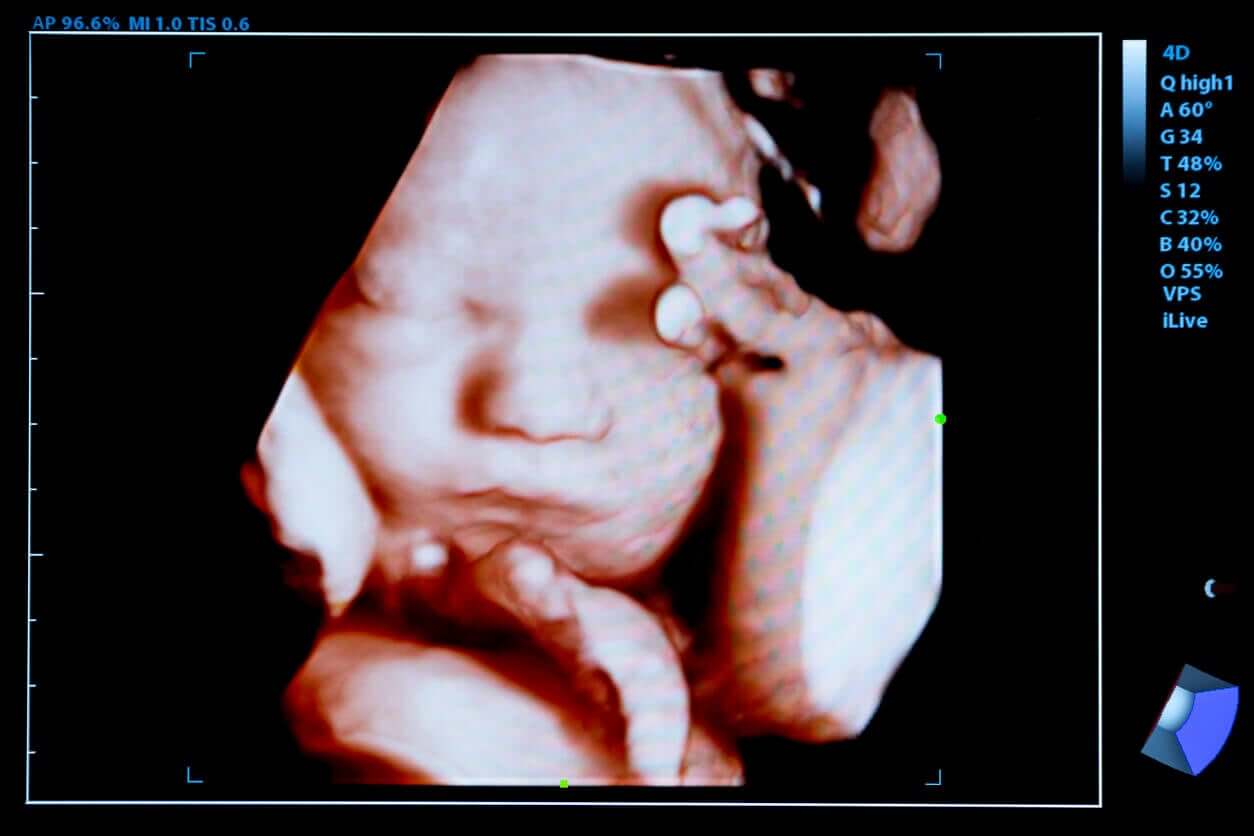
4D ultrasound is a technique by which consecutive images of the baby are obtained within the mother’s womb in a clearer way. To such an extent, that they allow you to observe the little one in motion like a video in real-time.
The usefulness of this technique is to visualize the fetal anatomy in greater detail and thus detect problems in its formation. And in addition, it allows you to discover your baby’s face for the first time.
Learn more about this radiological test and the benefits it offers.
What is 4D ultrasound?
Technology has revolutionized prenatal visits, especially with the evolution of ultrasound. Thanks to this, today, some treatable fetal complications can be diagnosed early.
4D ultrasound is a novel diagnostic test that allows detailed images of the fetus to be obtained through high-frequency ultrasound waves. Unlike the previous ones, it manages to reproduce around 24 3D images in one second and gives them movement.
Then, in addition to a detailed examination of the child’s body, you can see different movements that the child makes, such as smiling, thumb sucking, or yawning.
What is seen on 4D ultrasound?
Despite its specificity, this type of ultrasound doesn’t replace 2D ultrasound. The latter is more sensitive when it comes to confirming an early pregnancy, determining the number of babies, or gestational age.
Certainly, with 4D ultrasound, morphological alterations can be detailed in more advanced stages such as cleft lip, congenital heart disease, or baby skin problems. In addition to this, it’s useful to know the following aspects of pregnancy:
- Location and status of the placenta
- Fetal position
- Baby’s heart rate
- Sex of the baby
- Amount of amniotic fluid
- Interaction between babies in multiple pregnancies
- Fetal morphology
- Baby movements
When does the 4D ultrasound take place?
4D ultrasound can be performed at any time during pregnancy. However, in the first few months, the baby isn’t fully formed and the images may not be as clear. Even so, if you want to observe the baby’s sex at week 16, this technique can be implemented.
From the second trimester of gestation, especially between weeks 24 to 30, the characteristics of the baby are better observed and it’s the ideal time to perform this procedure. At this stage, the baby has an adequate size and there’s a greater amount of amniotic fluid, which allows the images to be better captured.
It’s important to note that after week 32, the baby grows larger, its position changes, and the amount of amniotic fluid decreases. This can make it difficult to see during the study and not show their features clearly.
How it is performed?
This test is performed by a radiologist or gynecologist in the office. Although it doesn’t require any preparation, the mother should be sufficiently hydrated and not use creams or oils on the abdomen on the day of the study.
To do it, the pregnant woman lies on a stretcher and uncovers her belly. The specialist places a small amount of cold gel on her belly and uses a device called a transducer to capture the baby’s images.
These moving images are projected on the screen and can also be recorded on CD to preserve the memory of that beautiful experience.
Advantages of 4D ultrasound

There are many advantages of this diagnostic test during pregnancy, especially in the emotional sense. Below, we list its benefits:
- It’s a harmless radiological test for mother and baby if done in the right place and in the right context.
- It’s painless and non-invasive.
- 4D ultrasound shows the baby’s movements and features quite clearly.
- It can detect some specific conditions such as cleft lip, palate, skin changes.
- It facilitates the cardiovascular evaluation of the baby.
- The procedure can identify traits of genetic syndromes, such as Down’s.
- It strengthens the emotional bond between the baby and their parents. By seeing the baby clearly, parents feel parenthood as something more real and exciting.
- The results are stored on CD and given to the parents.
It’s important to emphasize that 4D ultrasound doesn’t replace two-dimensional ultrasound, but rather complements it.
If you want to have a 4D ultrasound, you should consult with your doctor about the ideal date and make an appointment in advance.
4D ultrasound is a technique by which consecutive images of the baby are obtained within the mother’s womb in a clearer way. To such an extent, that they allow you to observe the little one in motion like a video in real-time.
The usefulness of this technique is to visualize the fetal anatomy in greater detail and thus detect problems in its formation. And in addition, it allows you to discover your baby’s face for the first time.
Learn more about this radiological test and the benefits it offers.
What is 4D ultrasound?
Technology has revolutionized prenatal visits, especially with the evolution of ultrasound. Thanks to this, today, some treatable fetal complications can be diagnosed early.
4D ultrasound is a novel diagnostic test that allows detailed images of the fetus to be obtained through high-frequency ultrasound waves. Unlike the previous ones, it manages to reproduce around 24 3D images in one second and gives them movement.
Then, in addition to a detailed examination of the child’s body, you can see different movements that the child makes, such as smiling, thumb sucking, or yawning.
What is seen on 4D ultrasound?
Despite its specificity, this type of ultrasound doesn’t replace 2D ultrasound. The latter is more sensitive when it comes to confirming an early pregnancy, determining the number of babies, or gestational age.
Certainly, with 4D ultrasound, morphological alterations can be detailed in more advanced stages such as cleft lip, congenital heart disease, or baby skin problems. In addition to this, it’s useful to know the following aspects of pregnancy:
- Location and status of the placenta
- Fetal position
- Baby’s heart rate
- Sex of the baby
- Amount of amniotic fluid
- Interaction between babies in multiple pregnancies
- Fetal morphology
- Baby movements
When does the 4D ultrasound take place?
4D ultrasound can be performed at any time during pregnancy. However, in the first few months, the baby isn’t fully formed and the images may not be as clear. Even so, if you want to observe the baby’s sex at week 16, this technique can be implemented.
From the second trimester of gestation, especially between weeks 24 to 30, the characteristics of the baby are better observed and it’s the ideal time to perform this procedure. At this stage, the baby has an adequate size and there’s a greater amount of amniotic fluid, which allows the images to be better captured.
It’s important to note that after week 32, the baby grows larger, its position changes, and the amount of amniotic fluid decreases. This can make it difficult to see during the study and not show their features clearly.
How it is performed?
This test is performed by a radiologist or gynecologist in the office. Although it doesn’t require any preparation, the mother should be sufficiently hydrated and not use creams or oils on the abdomen on the day of the study.
To do it, the pregnant woman lies on a stretcher and uncovers her belly. The specialist places a small amount of cold gel on her belly and uses a device called a transducer to capture the baby’s images.
These moving images are projected on the screen and can also be recorded on CD to preserve the memory of that beautiful experience.
Advantages of 4D ultrasound
There are many advantages of this diagnostic test during pregnancy, especially in the emotional sense. Below, we list its benefits:
- It’s a harmless radiological test for mother and baby if done in the right place and in the right context.
- It’s painless and non-invasive.
- 4D ultrasound shows the baby’s movements and features quite clearly.
- It can detect some specific conditions such as cleft lip, palate, skin changes.
- It facilitates the cardiovascular evaluation of the baby.
- The procedure can identify traits of genetic syndromes, such as Down’s.
- It strengthens the emotional bond between the baby and their parents. By seeing the baby clearly, parents feel parenthood as something more real and exciting.
- The results are stored on CD and given to the parents.
It’s important to emphasize that 4D ultrasound doesn’t replace two-dimensional ultrasound, but rather complements it.
If you want to have a 4D ultrasound, you should consult with your doctor about the ideal date and make an appointment in advance.
All cited sources were thoroughly reviewed by our team to ensure their quality, reliability, currency, and validity. The bibliography of this article was considered reliable and of academic or scientific accuracy.
- Cuadros, M. (2010). Ecografía 4D para el diagnóstico de malformaciones congénitas. Progresos de obstetricia y ginecología Vol. 53. Núm. 7 (Julio 2010).
- Lebit F. (2011). The Role of 4D Ultrasound in the Assessment of Fetal Behaviour. Maedica a journal of clinical medicine. 2011 Apr; 6(2): 120–127.
This text is provided for informational purposes only and does not replace consultation with a professional. If in doubt, consult your specialist.


