The Female Reproductive System: How It Works

The female reproductive system is the anatomical structure that makes fertilization, gestation and birth possible. The whole system is located in the pelvis area and includes organs that participate in reproduction.
Within the female reproductive system, there are external and internal organs. They are all interrelated and form a system in which each organ plays an important role.
The main functions of the reproductive system are: to produce reproductive cells, to allow for fertilization and to protect and nourish fertilized cells until they develop fully, culminating in the birth of a child.
External organs of the female reproductive system
The outer zone of the female reproductive system is called the “vulva” which means “cover.” Therefore, it’s located in the crotch and its main function is to protect the vaginal orifice and internal reproductive organs.
The external organs of the female reproductive system that are located in the vulva are the following:
- Mons pubis. This is a soft and fatty tissue on the pelvis that fulfills the role of protecting the internal organs. It starts to be covered by hair during puberty.
- External labia. These are two folds of skin in the shape of lips that limit the vulva externally. The space between the two is called the “vulvar cleft.” External labia therefore, prevent bacteria from entering the reproductive system.
- Internal labia. The internal labia are found inside the external labia and they’re similar in characteristics but a little smaller. They surround the vaginal orifice. In addition to being responsible for maintaining the vagina’s temperature, the internal labia also prevents the entry of foreign particles.
- Clitoris. The clitoris is an organ that is composed of 8,000 nerve endings. It starts in the internal labia and bifurcates into two corpora cavernosa inside the vagina. Its function is to generate pleasure during sexual intercourse.
- Urinary meatus. The urinary meatus is the duct through which urine comes out. It’s the outer section of the urethra and located between the clitoris and the vaginal orifice.
- Hymen. The hymen is a membrane that is located at the entrance of the vagina, therefore, it usually breaks during the first sexual intercourse or as a result of routine activities.
- Vulvar fork. The vulvar fork is where the external and internal labia meet.
- Perineum. It’s found between the pubis and the coccyx and its function is to protect the bladder, rectum and reproductive system.
Internal reproductive organs
The following are the internal organs of the female reproductive system:
- Vagina. The vagina is muscular tissue in the form of a tube; it connects the uterus with the exterior. It’s elastic and ends in a hole called the vaginal orifice. It’s the connection between the vulva and the internal reproductive organs.
- Uterus. The uterus is a hollow muscular organ. It’s located between the vagina and the Fallopian tubes. The uterus is where gestation occurs. It’s made up of three layers: the endometrium, smooth muscles and elastic tissue.
- Fallopian tubes. These organs are shaped like tubes and they are the connection between the uterus and the ovaries. Their function is to send the ovules or reproductive cells from the ovary to the uterus.
- Ovaries. The ovaries are two organs and they are approximately the size of an almond. Their function is to produce an egg or a reproductive cell every 28 days. Furthermore the ovaries also produce sex hormones.
The operation of the system
At birth, a woman has hundreds of thousands of oocytes or reproductive cells inside her ovaries. Due to the production of sex hormones, the menstrual cycle begins during puberty.
So, after puberty, one oocyte is released every month. If an oocyte is fertilized, it remains in the uterus and the formation of a new being begins.
If the oocyte isn’t fertilized, it dries and leaves the body about two weeks after it was released from the ovaries. It exits the uterus along with blood and the internal lining of this organ.
This process is known as menstruation. The first time it occurs is called menarche.
When a sperm fertilizes an oocyte, a zygote is formed. The zygote then develops into a blastocyst which is a ball of cells with fluid inside. It nests in the endometrium which is one of the layers of the uterus.
In addition, sex hormones are important in the consolidation of the process. The final result is implantation.
Finally, the embryo is formed which will then develop into a fetus.
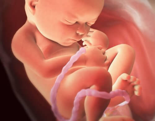
The fetus then floats in amniotic fluid. In addition, the fetus breathes and is fed through the placenta. The placenta is able to communicate with the fetus thanks to the umbilical cord.
After nine months of gestation, the cervix dilates and the uterus walls contract and push the baby towards the exterior. That’s how a baby is born.
The female reproductive system is the anatomical structure that makes fertilization, gestation and birth possible. The whole system is located in the pelvis area and includes organs that participate in reproduction.
Within the female reproductive system, there are external and internal organs. They are all interrelated and form a system in which each organ plays an important role.
The main functions of the reproductive system are: to produce reproductive cells, to allow for fertilization and to protect and nourish fertilized cells until they develop fully, culminating in the birth of a child.
External organs of the female reproductive system
The outer zone of the female reproductive system is called the “vulva” which means “cover.” Therefore, it’s located in the crotch and its main function is to protect the vaginal orifice and internal reproductive organs.
The external organs of the female reproductive system that are located in the vulva are the following:
- Mons pubis. This is a soft and fatty tissue on the pelvis that fulfills the role of protecting the internal organs. It starts to be covered by hair during puberty.
- External labia. These are two folds of skin in the shape of lips that limit the vulva externally. The space between the two is called the “vulvar cleft.” External labia therefore, prevent bacteria from entering the reproductive system.
- Internal labia. The internal labia are found inside the external labia and they’re similar in characteristics but a little smaller. They surround the vaginal orifice. In addition to being responsible for maintaining the vagina’s temperature, the internal labia also prevents the entry of foreign particles.
- Clitoris. The clitoris is an organ that is composed of 8,000 nerve endings. It starts in the internal labia and bifurcates into two corpora cavernosa inside the vagina. Its function is to generate pleasure during sexual intercourse.
- Urinary meatus. The urinary meatus is the duct through which urine comes out. It’s the outer section of the urethra and located between the clitoris and the vaginal orifice.
- Hymen. The hymen is a membrane that is located at the entrance of the vagina, therefore, it usually breaks during the first sexual intercourse or as a result of routine activities.
- Vulvar fork. The vulvar fork is where the external and internal labia meet.
- Perineum. It’s found between the pubis and the coccyx and its function is to protect the bladder, rectum and reproductive system.
Internal reproductive organs
The following are the internal organs of the female reproductive system:
- Vagina. The vagina is muscular tissue in the form of a tube; it connects the uterus with the exterior. It’s elastic and ends in a hole called the vaginal orifice. It’s the connection between the vulva and the internal reproductive organs.
- Uterus. The uterus is a hollow muscular organ. It’s located between the vagina and the Fallopian tubes. The uterus is where gestation occurs. It’s made up of three layers: the endometrium, smooth muscles and elastic tissue.
- Fallopian tubes. These organs are shaped like tubes and they are the connection between the uterus and the ovaries. Their function is to send the ovules or reproductive cells from the ovary to the uterus.
- Ovaries. The ovaries are two organs and they are approximately the size of an almond. Their function is to produce an egg or a reproductive cell every 28 days. Furthermore the ovaries also produce sex hormones.
The operation of the system
At birth, a woman has hundreds of thousands of oocytes or reproductive cells inside her ovaries. Due to the production of sex hormones, the menstrual cycle begins during puberty.
So, after puberty, one oocyte is released every month. If an oocyte is fertilized, it remains in the uterus and the formation of a new being begins.
If the oocyte isn’t fertilized, it dries and leaves the body about two weeks after it was released from the ovaries. It exits the uterus along with blood and the internal lining of this organ.
This process is known as menstruation. The first time it occurs is called menarche.
When a sperm fertilizes an oocyte, a zygote is formed. The zygote then develops into a blastocyst which is a ball of cells with fluid inside. It nests in the endometrium which is one of the layers of the uterus.
In addition, sex hormones are important in the consolidation of the process. The final result is implantation.
Finally, the embryo is formed which will then develop into a fetus.
The fetus then floats in amniotic fluid. In addition, the fetus breathes and is fed through the placenta. The placenta is able to communicate with the fetus thanks to the umbilical cord.
After nine months of gestation, the cervix dilates and the uterus walls contract and push the baby towards the exterior. That’s how a baby is born.
This text is provided for informational purposes only and does not replace consultation with a professional. If in doubt, consult your specialist.


