Infant Hemangiomas: Common Tumors in Infants

Infant hemangiomas are benign tumors caused by an exaggerated or uncontrolled growth of blood vessels. They appear on the baby’s body a few days or weeks after birth.
Hemangiomas develop quickly in the first few months of life. They tend to go into spontaneous remission and to disappear between 5 and 7 years of age. Nonetheless, there are some rare cases where complications may arise and they may need medical treatment.
In general, this type of tumor is varied in size. They can appear at any time during the first three months of life and they continue to grow once they appear, and later stabilize during the first year. If the hemangioma is large, it can continue to grow throughout the entire first year. In any case, they will only start to diminish in the child’s third year and they will continue to fade slowly until they disappear. Sometimes they leave a slight scar or other traces of their former presence that is more or less noticeable on the skin.
Treatment is only necessary if the tumor is inhibiting the functioning of an organ or the scars pose a real esthetic problem for the child. The lesions start to slowly dissolve beginning at about 12 to 18 months. The majority of hemangiomas remain localized to the skin and do not require any medical attention.
What are the types of infant hemangiomas?
Infant hemangiomas are classified based on their appearance. The section below describes the three major types of infant hemangiomas:
Superficial infant hemangioma
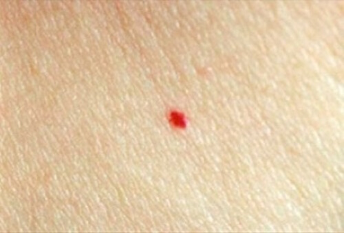
This presents as a scarlet red lesion when it first manifests, with a clean border and an irregular surface. This is the most common type of hemangioma and it appears in the first three weeks of life.
Deep or subcutaneous infant hemangioma:
These nodules have a bluish tinge. This type is less common and appears later, at around three or four months old.
Mixed infant hemangioma:
This affects the dermis and hypodermis. It has a superficial red component that stands out with a bluish halo.
How do hemangiomas develop
The characteristic evolution of this kind of tumor occurs in three phases. In the first proliferative phase, the tumor grows rapidly in size and continues to grow until the child is between 6 and 12 months old. The cutaneous part of the lesion is bright red and elevated with respect to the healthy skin surrounding it. When the hemangioma is strictly subcutaneous, it usually presents some light swelling with a bluish tinge. Sometimes it’s possible to perceive the drainage veins in the lesion.
The first phase is followed by a second phase where the hemangioma stops growing. This is the phase when the tumor begins to reduce in size. Clinically, the surface of the skin becomes paler. This stage tends to last from 18 months to 7 years of age. It depends on how much subcutaneous tissue the hemangioma involved.
Finally, the last stage is when the lesion disappears. At this stage, there can be residual fibroadipose tissue associated or not with the presence of spider veins.
How should infant hemangiomas be treated?
What is certain is that the majority of small hemangiomas do not require any special treatment. Nonetheless, it’s necessary to monitor hemangiomas that appear on the face, even superficial ones because their location and evolution can cause functional problems for the child.
The ideal treatment will depend on factors that are specific to the patient and that should be personalized depending on the location, size, and severity of the lesions. Local treatments and wound care is essential if the tumor presents with any ulceration and parents should let the doctors know if the child experiences any bleeding or pain.
The treatments recommended for those hemangiomas requiring care include corticosteroids, cryotherapy, laser therapy, and more rarely surgical interventions. In the absence of factors that complicate the prognosis or that cause damage to vital organs, doctors usually recommend avoiding surgery or other aggressive interventions because of the risk of scaring.
Finally, remember that hemangiomas represent one of the most common infant tumors. Researchers are still not clear on the origins of these tumors or what causes the uncontrolled growth of blood vessels. Hopefully, there will be more research to better understand them in the future.
Clearly, there’s no need to worry excessively if your baby happens to develop a hemangioma. With a little patience, they tend to resolve all on their own. It’s important the doctor determines if there are any complications, so they can be addressed in a timely way that reduces any potential scaring.
Infant hemangiomas are benign tumors caused by an exaggerated or uncontrolled growth of blood vessels. They appear on the baby’s body a few days or weeks after birth.
Hemangiomas develop quickly in the first few months of life. They tend to go into spontaneous remission and to disappear between 5 and 7 years of age. Nonetheless, there are some rare cases where complications may arise and they may need medical treatment.
In general, this type of tumor is varied in size. They can appear at any time during the first three months of life and they continue to grow once they appear, and later stabilize during the first year. If the hemangioma is large, it can continue to grow throughout the entire first year. In any case, they will only start to diminish in the child’s third year and they will continue to fade slowly until they disappear. Sometimes they leave a slight scar or other traces of their former presence that is more or less noticeable on the skin.
Treatment is only necessary if the tumor is inhibiting the functioning of an organ or the scars pose a real esthetic problem for the child. The lesions start to slowly dissolve beginning at about 12 to 18 months. The majority of hemangiomas remain localized to the skin and do not require any medical attention.
What are the types of infant hemangiomas?
Infant hemangiomas are classified based on their appearance. The section below describes the three major types of infant hemangiomas:
Superficial infant hemangioma
This presents as a scarlet red lesion when it first manifests, with a clean border and an irregular surface. This is the most common type of hemangioma and it appears in the first three weeks of life.
Deep or subcutaneous infant hemangioma:
These nodules have a bluish tinge. This type is less common and appears later, at around three or four months old.
Mixed infant hemangioma:
This affects the dermis and hypodermis. It has a superficial red component that stands out with a bluish halo.
How do hemangiomas develop
The characteristic evolution of this kind of tumor occurs in three phases. In the first proliferative phase, the tumor grows rapidly in size and continues to grow until the child is between 6 and 12 months old. The cutaneous part of the lesion is bright red and elevated with respect to the healthy skin surrounding it. When the hemangioma is strictly subcutaneous, it usually presents some light swelling with a bluish tinge. Sometimes it’s possible to perceive the drainage veins in the lesion.
The first phase is followed by a second phase where the hemangioma stops growing. This is the phase when the tumor begins to reduce in size. Clinically, the surface of the skin becomes paler. This stage tends to last from 18 months to 7 years of age. It depends on how much subcutaneous tissue the hemangioma involved.
Finally, the last stage is when the lesion disappears. At this stage, there can be residual fibroadipose tissue associated or not with the presence of spider veins.
How should infant hemangiomas be treated?
What is certain is that the majority of small hemangiomas do not require any special treatment. Nonetheless, it’s necessary to monitor hemangiomas that appear on the face, even superficial ones because their location and evolution can cause functional problems for the child.
The ideal treatment will depend on factors that are specific to the patient and that should be personalized depending on the location, size, and severity of the lesions. Local treatments and wound care is essential if the tumor presents with any ulceration and parents should let the doctors know if the child experiences any bleeding or pain.
The treatments recommended for those hemangiomas requiring care include corticosteroids, cryotherapy, laser therapy, and more rarely surgical interventions. In the absence of factors that complicate the prognosis or that cause damage to vital organs, doctors usually recommend avoiding surgery or other aggressive interventions because of the risk of scaring.
Finally, remember that hemangiomas represent one of the most common infant tumors. Researchers are still not clear on the origins of these tumors or what causes the uncontrolled growth of blood vessels. Hopefully, there will be more research to better understand them in the future.
Clearly, there’s no need to worry excessively if your baby happens to develop a hemangioma. With a little patience, they tend to resolve all on their own. It’s important the doctor determines if there are any complications, so they can be addressed in a timely way that reduces any potential scaring.
All cited sources were thoroughly reviewed by our team to ensure their quality, reliability, currency, and validity. The bibliography of this article was considered reliable and of academic or scientific accuracy.
- Baselga Torres, E.; Bernabéu Wittel, J.; van Esso Arbolave, D. L., et al. (2016). “Consenso español sobre el hemangioma infantil”, Anales de Pediatría, 85 (5): 256-265.
- Carvalho, S.; Machado, S., & Selores, M. (2016). “Hemangioma infantil e propranolol oral: recomendações atuais TT”, Nascer e Crescer, 25 (3): 154-158.
- Huraki, P. Y., & Goldenberg, D. C. (2010). “Diagnóstico e tratamento do hemangioma infantil”, Rev. Bras. Cir. Plást, 25(2): 388-397.
This text is provided for informational purposes only and does not replace consultation with a professional. If in doubt, consult your specialist.


