Dental Cysts in Children: How to Treat Them
Infections in dental pieces that aren’t treated in a timely manner can give rise to the formation of dental cysts in children. Although this is a benign disease, its presence in the mouth of children usually worries and concerns parents.
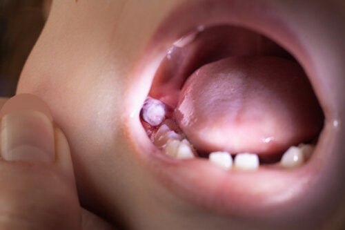
This complication can manifest itself as a lump in the gum, which produces inflammation of the area and pain. But other times, it can go unnoticed and only a dentist can detect it during check-ups.
Dental cysts in children shouldn’t be overlooked, as they can lead to other health problems. In this article, we’ll tell you everything you need to know about them so that you know how to act.
What are dental cysts in children?
A dental cyst is a lesion that develops inside the maxillary bones. Its shape is that of a closed pocket with its own membrane, which surrounds a collection (of liquid, cells, or air) and which develops abnormally.
The walls of the cyst are formed by cells, which divide, grow, and increase the volume of the cavity. They gradually destroy the vicinity of the bone in the area where they’re located, and if not treated in a timely manner, can compromise bone health. In addition, the fluid contained in these lesions often becomes infected by bacteria in the mouth and constitutes the focus of a deep infection.
In general, the dental cyst in children is considered a non-tumorous and benign lesion. But in some situations, it can give rise to malignant processes that develop accordingly.
Symptoms of dental cysts in children
The growth of a dental cyst in the mouth of children is usually slow and often asymptomatic. The manifestations appear when the liquid contained in this lesion becomes infected or when the cyst reaches a considerable size to deform the surrounding bone.
Here are some of the manifestations of dental cysts in children when they’re symptomatic:
- Pain in the gum
- Swelling or a lump in the area of the cyst
- Fistulas that drain fluid through the gum (to the inside of the mouth or to the outside)
- Mobility and displacement of teeth near the lesion
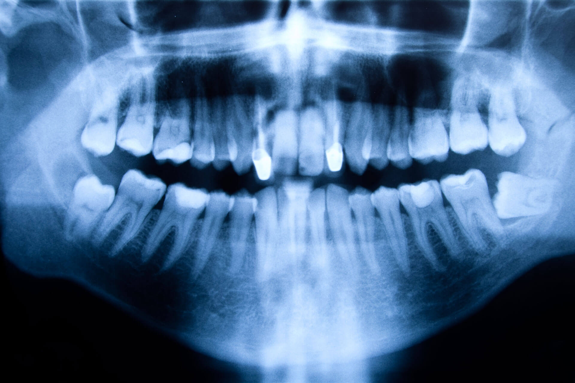
- Resorption of the roots of the teeth near the cyst (this is observed with dental radiographs)
- Paresthesias (tingling) or alterations of the sensitivity of the lips, mouth, or face due to compression of local nerve structures
The causes of dental cysts
Dental cysts in children are considered to be of dental origin and are therefore called odontogenic cysts. The epithelial cells that cover them come from the embryonic period and are trapped inside the bone. They develop spontaneously or after an infection in nearby teeth.
The radicular dental cyst is of inflammatory origin and is the most frequent type in children. It’s located at the tip of the root of an infected tooth, for example, deep cavities or trauma that resulted in pulp necrosis.
The dead and infected pulp causes the accumulation of pus and inflammation of the root of the tooth. With this, the remains of embryonic cells are activated and a cavity with pus develops inside the maxillary bone. This is the root cyst.
Developmental cysts can also form in children, the origin of which is not associated with inflammatory stimuli. The most common example is the dentigerous cyst. This cavity forms around teeth that haven’t yet erupted and are common in the areas of the wisdom teeth or canines during adolescence. However, they can affect any dental element.
The treatment of dental cysts in children
The first step in the proper treatment of dental cysts in children is to have an accurate diagnosis. And for this, regular dental visits are essential.
In some cases, the dental cyst may present symptoms that lead parents to seek an urgent solution. But other times, these lesions are detected as clinical or radiographic findings on routine dental exams.
When a cyst is suspected, the dentist will take X-rays of the mouth to determine the exact location, size, condition of the tooth causing the cyst, and nearby structures involved. Sometimes, other complementary tests may also be requested, such as puncture or aspiration of the cyst to study the nature of its cells under the microscope.
As for treatment, it’s sometimes possible to try conservative treatment, avoiding the extraction of the problematic tooth element. For this, first, the tooth is endodontically treated and then antibiotic treatment is implemented to cure the infection.
Then, the evolution of the clinical picture is controlled through X-rays and if the lesion persists, a small surgery is performed to eliminate the cyst definitively.
In particular cases, the extent of the process or damage to the tooth element merits the extraction of the tooth in question and the cyst together. This surgery allows the removal of all the infectious tissue that could be located inside the bone. If the bone damage has been very significant, it may be necessary to carry out treatments to regenerate this tissue.
Once the cyst has been removed, it’s essential to analyze its contents under the microscope to evaluate the type of lesion in detail. And in the postoperative period, it’s crucial to follow an antibiotic regimen to protect the wound from possible opportunistic infections by germs in the mouth.

The prevention of dental cysts in children
The most frequent cause of the appearance of dental cysts in children is the infection of the pulp. Therefore, the best way to prevent the appearance of these lesions is to avoid diseases that cause the death of the nerve, such as cavities.
With proper dental hygiene, a healthy diet, and regular visits to the pediatric dentist, it’s possible to maintain the oral health of children. And if the dentist detects that something is wrong, early treatment can be carried out to avoid the development of more complicated consequences, such as the appearance of cysts.
If your child has pain or swelling in the mouth, you should go to the dentist immediately. As we mentioned, these types of lesions grow and spread over time. And in these cases, the treatments required are more invasive and uncomfortable.
With proper dental care and controls, you can avoid dental cysts in your child. And if they do appear, they should be treated and stopped as soon as they’re detected..
All cited sources were thoroughly reviewed by our team to ensure their quality, reliability, currency, and validity. The bibliography of this article was considered reliable and of academic or scientific accuracy.
- Álvarez, J. D. P. (2014). Quiste radicular de origen odontogénico. Revista Nacional de Odontología, 10(19), 91-100.
- Pulido Valladares, Y., Torres Rodríguez, L. E., & Gounelas Amat, S. (2019). Quiste dentígero en la atención pediátrica multidisciplinaria. Revista de Ciencias Médicas de Pinar del Río, 23(3), 473-479.
- Pina Godoy, G., Dantas da Silveira, É. J., Gordón-Núñez, M. A., Guedes Queiroz, L. M., & Medeiros Dantas Gomes, D. (2007). Quistes de los maxilares en niños: un análisis clínico. Acta Odontológica Venezolana, 45(4), 546-549.
- Verbel Bohórquez, J., Ramos Manotas, J., & Díaz Caballero, A. (2015). Radiografía periapical como herramienta en el diagnóstico y tratamiento de quiste periapical. Avances en Odontoestomatología, 31(1), 25-29.
- de la Rosa Ricardo, L., Rodríguez, K. L. P., & Simeón-Pérez, R. E. (2021). Alternativa de tratamiento endodóntico ante procesos periapicales. Reporte de caso. 16 de Abril, 60(279), 1-5.
- Arredondo, D. O. D., Cedeño, J. M. O., Torres, I. L., & Hinojosa, H. C. (2018). Manejo multidisciplinario de quiste periapical. Reporte de caso. Revista Mexicana de Estomatología, 5(2), 14-17.
- Rodríguez Castellanos, A., Barrera Garcell, M., & Rodríguez Rey, H. M. (2021). Tratamiento multidisciplinario en un niño afectado por un quiste dentígero. Medisan, 25(4), 924-933.
- Pérez López, G., Soto Fernández, Á., Jequin Savariego, E., López Hernández, A., & Villalonga Pérez, G. (2003). Quiste odontógeno: Presentación de caso. Revista Cubana de Estomatología, 40(3), 0-0.