What the Fetal Heartbeat in the Mother's Womb is Like

Written and verified by the nurse Leidy Mora Molina
One of the first parameters that a specialist evaluates when confirming a pregnancy through ultrasound is the fetal heartbeat in the mother’s womb. Listening to it becomes one of the most emotional moments, as this way to verify that there’s a baby forming inside her
These heartbeats can be heard only after the sixth week of pregnancy, generally in the first ultrasound. But what are they like? Is their frequency fast or slow? Do they change during gestation? Find out below what a fetal heartbeat is like, how it evolves in the mother’s womb, and how cardiac well-being is assessed at this stage.
The fetal heartbeat
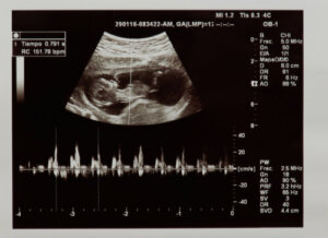
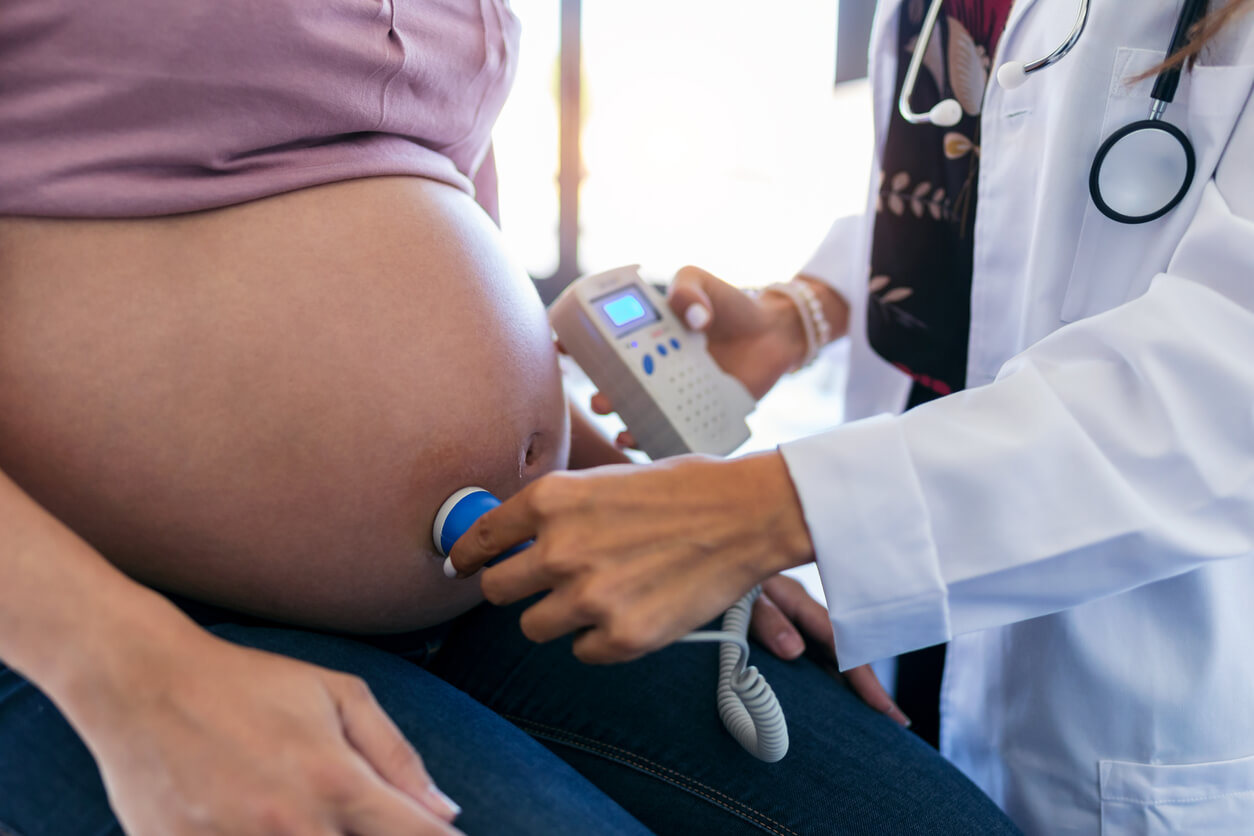
By heartbeat, we’re referring to the number of times the heart contracts during one minute. The fetal heart rate is identifiable in the first weeks of gestation by means of an ultrasound or a monitor known as fetal Doppler.
First, the ultrasound technician will assess whether the heartbeat is actually present, thus checking fetal vitality. In addition, they’ll evaluate how many times the heart beats during one minute. The values considered normal in the fetal stage are between 120 and 160 beats per minute.
Heart rate problems
A heart rate outside normal parameters is an indication that something’s wrong with the pregnancy. It may be related to a lack of oxygen supply to the baby or to other problems, such as heart malformations.
Based on this evaluation, the specialist will then determine if there’s a risk of miscarriage. In advanced pregnancies, the need for labor induction or cesarean section may be assessed, according to the gestational age of the baby.
In any case, it should be kept in mind that during the second trimester, the heart’s electrical conduction pathways begin to develop and mature, which may cause the heart rate to alter momentarily. This is physiological and is not a cause for alarm, but in case the doctor detects that something is not right, he will have to carry out continuous evaluations.
Slow fetal heart rate
Occasionally, during the first trimester, a slower fetal heart rate is observed. Although this is considered normal (especially in the first weeks), some studies associate it with an increased risk of miscarriage.
In this regard, the result of a study carried out in Chile in 2002 was that the risk of reproductive loss at six and seven weeks of gestation increases 19 times when the embryonic heart rate is less than 90 beats per minute, while it’s seven times higher when it’s between 90 and 100.
This is also confirmed by a study carried out by researchers at Cornell University, published in the journal Fertility and Sterility. In this case, they concluded that a decrease in the embryonic heart rate in the first trimester, independently of maternal age, can help predict the prognosis of pregnancy in the first trimester after in vitro fertilization.
Going to the doctor will be key so that the professional can assess whether pregnancy can be achieved or if there are other problems such as chromosomal alterations or fetal malformations that prevent it.
Evolution of the fetal heartbeat in the mother’s womb
Now, to understand if the fetal heart rate is normal or if there’s a fetal arrhythmia, it’s important to know what the evolution of the fetal heartbeat is like during gestation.
In this regard, between the fifth and sixth week of gestation, the heart gives its first beats. At this time, the heart rate is slow and is considered normal at around 80 beats per minute.
From this point, the heart rate begins to increase. It’s estimated that it increases by about 3 beats per minute daily until it reaches an average of 175, which occurs around the ninth week. Once these values are reached, it tends to fall slightly to stabilize between 120 and 160 and remain this way throughout gestation.
Can the baby’s heart be assessed in the mother’s womb?
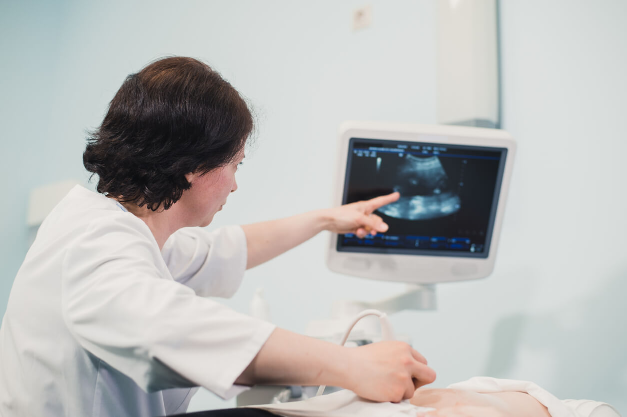
Assessment of the anatomy and physiology of the baby’s heart can be performed in the mother’s womb. This is thanks to the ultrasounds performed during prenatal check-ups, which make it possible to assess, among other parameters, cardiac health, and the fetal heartbeat.
Certainly, during the second trimester, morphological ultrasound is indicated, where the baby’s organs are studied in detail. Here, the cardiac structures of the fetus, such as the chambers, valves, and blood vessels of the heart, are carefully observed.
It also evaluates the functioning of the heart and whether there are any problems in its mechanism or conduction. Morphological ultrasound also diagnoses the existence of congenital heart disease that may cause problems after the baby is born.
One of the first parameters that a specialist evaluates when confirming a pregnancy through ultrasound is the fetal heartbeat in the mother’s womb. Listening to it becomes one of the most emotional moments, as this way to verify that there’s a baby forming inside her
These heartbeats can be heard only after the sixth week of pregnancy, generally in the first ultrasound. But what are they like? Is their frequency fast or slow? Do they change during gestation? Find out below what a fetal heartbeat is like, how it evolves in the mother’s womb, and how cardiac well-being is assessed at this stage.
The fetal heartbeat
By heartbeat, we’re referring to the number of times the heart contracts during one minute. The fetal heart rate is identifiable in the first weeks of gestation by means of an ultrasound or a monitor known as fetal Doppler.
First, the ultrasound technician will assess whether the heartbeat is actually present, thus checking fetal vitality. In addition, they’ll evaluate how many times the heart beats during one minute. The values considered normal in the fetal stage are between 120 and 160 beats per minute.
Heart rate problems
A heart rate outside normal parameters is an indication that something’s wrong with the pregnancy. It may be related to a lack of oxygen supply to the baby or to other problems, such as heart malformations.
Based on this evaluation, the specialist will then determine if there’s a risk of miscarriage. In advanced pregnancies, the need for labor induction or cesarean section may be assessed, according to the gestational age of the baby.
In any case, it should be kept in mind that during the second trimester, the heart’s electrical conduction pathways begin to develop and mature, which may cause the heart rate to alter momentarily. This is physiological and is not a cause for alarm, but in case the doctor detects that something is not right, he will have to carry out continuous evaluations.
Slow fetal heart rate
Occasionally, during the first trimester, a slower fetal heart rate is observed. Although this is considered normal (especially in the first weeks), some studies associate it with an increased risk of miscarriage.
In this regard, the result of a study carried out in Chile in 2002 was that the risk of reproductive loss at six and seven weeks of gestation increases 19 times when the embryonic heart rate is less than 90 beats per minute, while it’s seven times higher when it’s between 90 and 100.
This is also confirmed by a study carried out by researchers at Cornell University, published in the journal Fertility and Sterility. In this case, they concluded that a decrease in the embryonic heart rate in the first trimester, independently of maternal age, can help predict the prognosis of pregnancy in the first trimester after in vitro fertilization.
Going to the doctor will be key so that the professional can assess whether pregnancy can be achieved or if there are other problems such as chromosomal alterations or fetal malformations that prevent it.
Evolution of the fetal heartbeat in the mother’s womb
Now, to understand if the fetal heart rate is normal or if there’s a fetal arrhythmia, it’s important to know what the evolution of the fetal heartbeat is like during gestation.
In this regard, between the fifth and sixth week of gestation, the heart gives its first beats. At this time, the heart rate is slow and is considered normal at around 80 beats per minute.
From this point, the heart rate begins to increase. It’s estimated that it increases by about 3 beats per minute daily until it reaches an average of 175, which occurs around the ninth week. Once these values are reached, it tends to fall slightly to stabilize between 120 and 160 and remain this way throughout gestation.
Can the baby’s heart be assessed in the mother’s womb?
Assessment of the anatomy and physiology of the baby’s heart can be performed in the mother’s womb. This is thanks to the ultrasounds performed during prenatal check-ups, which make it possible to assess, among other parameters, cardiac health, and the fetal heartbeat.
Certainly, during the second trimester, morphological ultrasound is indicated, where the baby’s organs are studied in detail. Here, the cardiac structures of the fetus, such as the chambers, valves, and blood vessels of the heart, are carefully observed.
It also evaluates the functioning of the heart and whether there are any problems in its mechanism or conduction. Morphological ultrasound also diagnoses the existence of congenital heart disease that may cause problems after the baby is born.
All cited sources were thoroughly reviewed by our team to ensure their quality, reliability, currency, and validity. The bibliography of this article was considered reliable and of academic or scientific accuracy.
- American pregnacy association (2022). Arritmia fetal. Recuperado de: https://americanpregnancy.org/es/healthy-pregnancy/pregnancy-complications/fetal-arrhythmia/
- Muñoz, H. et al (2002). Frecuencia cardiaca embrionaria y riesgo de pérdida reproductiva. Revista chilena de obstetricia y ginecología v.67 n.6 Santiago 2002. Recuperado de: https://www.scielo.cl/scielo.php?script=sci_arttext&pid=S0717-75262002000600001
- Rauch, E. et al (2008). Embryonic heart rate as a predictor of first-trimester pregnancy loss in infertility patients after in vitro fertilization. Fertility and sterility. Vol. 91, N° 6, pp. 2451-2454. Recuperado de: https://www.fertstert.org/article/S0015-0282(08)00597-9/fulltext
- Romero, G. et al (2003). Valores normales de la frecuencia cardíaca fetal. Clínica e Investigación en Ginecología y Obstetricia. Vol. 30. Núm. 9. Pp. 293-298. Recuperado de: https://www.elsevier.es/es-revista-clinica-e-investigacion-ginecologia-obstetricia-7-articulo-valores-normales-frecuencia-cardiaca-fetal-13055007
This text is provided for informational purposes only and does not replace consultation with a professional. If in doubt, consult your specialist.


