Morphology Scan: Everything You Need to Know

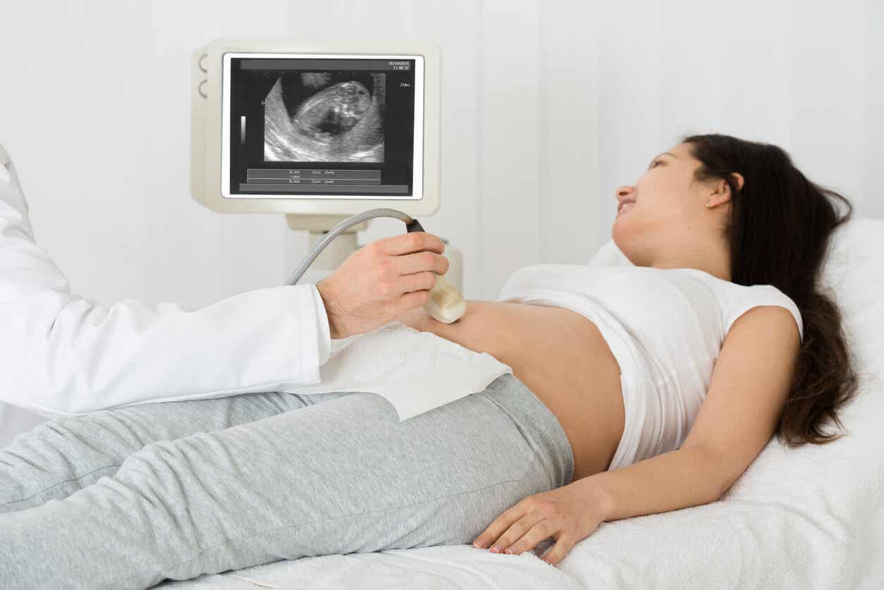
Did you know that during your 20th week of pregnancy doctors can identify whether your baby suffers from heart problems or other defects? This is possible by taking a morphology scan, a test that allows professionals to see the baby’s organs and tissues, and other structures, like the placenta.
Morphology scans provide important data about your baby, such as malformations or other defects that can cause problems over the course of the pregnancy. In this article, we’ll talk about this test and why it’s so important to take it during the second trimester.
Morphology scan: What is it?
Morphology scans, also known as ultrasound scans, use high-frequency sound waves to transmit two-dimensional or three-dimensional images of the uterus. By doing this, it’s possible to obtain images of the babies and the structural characteristics of their organs and body systems.

Specialists recommend this test to monitor if babies’ tissues have developed properly. Furthermore, they get to see if these tissues are growing as expected. What’s more, this scan evaluates the location of the placenta, the umbilical cord, and the amount of amniotic fluid present in the cavity.
Why do pregnant women have to wait until the 20th week of pregnancy to take this test?
Morphology scans are carried out during the second trimester of pregnancy, especially between the 20th and the 22nd week. This is because, during these weeks, babies have formed their systems and organs almost completely. Therefore, it’s possible to evaluate their body structures, such as their heart valves.
What’s more, during the 20th week of pregnancy, there’s more amniotic fluid around the baby. As a result, the images obtained are clearer so the specialist can evaluate them better.
What can you see in a morphology scan?
This ultrasound evaluates the general aspects of your pregnancy and your baby’s anatomy. Let’s see what specialists can identify when performing a morphology scan:
General aspects of your pregnancy
- Gestational age: Depending on the babies’ measurements, specialists identify the gestational age of the pregnancy. In order to do this, they measure babies’ length, the size of their head, their femur, and the leg bone.
- The number of babies: Specialists confirm the number of babies in the uterus. What’s more, if it’s possible, they evaluate internal structures, such as the placenta and the yolk sac. This way, they’ll find out whether babies are fraternal or identical twins.
- Fetal vitality: They also check if babies are active by analyzing their movements. Furthermore, they see if babies’ heart rate is correct.
- The amount of amniotic fluid.
- Placental position.
- Umbilical cord: Specialists assess the number of blood vessels (one vein, two arteries).
Babies’ anatomic aspects
Specialists analyze babies’ anatomy by looking at their structures and organs.
- Fetal biometry: Specialists analyze the parietal bones of the skull, the head, and the circumference of the abdomen. They also check the size of the femur, the babies’ estimated weight, and their percentile to see if their size is comparable to those of fetuses from the same gestational age.
- Bone formation in the head and the column, development of the central nervous system, and other structures, such as the eyes, palate, and lips.
- Specialists evaluate the position of the lungs and heart. Furthermore, they check blood vessels and valves.
- They also analyze the organs involved in the gastrointestinal and renal systems. In addition, specialists check the umbilical cord and the abdomen.
- The length and symmetry of the upper and lower limbs.
- Finally, what most parents desire the most: Doctors are able to discover the sex of the baby.
The importance of morphology scans during pregnancy
Morphology scans also help specialists diagnose the following aspects:
- Congenital malformations: These can occur in 2 % of pregnancies. Some examples of these malformations are spina bifida, congenital heart diseases, like tetralogy of Fallot, cleft lip, among others.
- Abnormalities in the placenta position, allowing specialists to identify placenta previa.
- If the baby has the right amount of amniotic fluid.
- If there are any chromosomal abnormalities, like Down Syndrome.
Certainty in the results of this test
The data obtained from the morphology scan are the results of specialists’ direct observations. Therefore, they’re reliable. When it’s not possible to observe clear images, specialists repeat this test a few weeks later. Furthermore, certain other tests can back up the results of the morphology scan.
How are morphology scans carried out?
First, you must bear in mind that it may take approximately 45 minutes. You can go with your partner, so both of you get the chance to see the baby and listen to what the doctor has to say.
- There’s no need to go with a full bladder. However, it could help the doctor get a better look at the images.
- The doctor will tell you to lay down and relax for a few minutes.
- Morphology scans usually take around 30 minutes. However, they can take a bit longer, too.
- This test bears no risk for the mother or the baby. It doesn’t cause any pain, either.
Finally, it’s important to know that the morphology scan is one of the most important tests you should take during pregnancy. By taking it, you’ll be able to identify possible malformations or risks that could complicate the pregnancy. Therefore, remember to have one during your 20th week of pregnancy, and follow your doctor’s instructions.
Did you know that during your 20th week of pregnancy doctors can identify whether your baby suffers from heart problems or other defects? This is possible by taking a morphology scan, a test that allows professionals to see the baby’s organs and tissues, and other structures, like the placenta.
Morphology scans provide important data about your baby, such as malformations or other defects that can cause problems over the course of the pregnancy. In this article, we’ll talk about this test and why it’s so important to take it during the second trimester.
Morphology scan: What is it?
Morphology scans, also known as ultrasound scans, use high-frequency sound waves to transmit two-dimensional or three-dimensional images of the uterus. By doing this, it’s possible to obtain images of the babies and the structural characteristics of their organs and body systems.
Specialists recommend this test to monitor if babies’ tissues have developed properly. Furthermore, they get to see if these tissues are growing as expected. What’s more, this scan evaluates the location of the placenta, the umbilical cord, and the amount of amniotic fluid present in the cavity.
Why do pregnant women have to wait until the 20th week of pregnancy to take this test?
Morphology scans are carried out during the second trimester of pregnancy, especially between the 20th and the 22nd week. This is because, during these weeks, babies have formed their systems and organs almost completely. Therefore, it’s possible to evaluate their body structures, such as their heart valves.
What’s more, during the 20th week of pregnancy, there’s more amniotic fluid around the baby. As a result, the images obtained are clearer so the specialist can evaluate them better.
What can you see in a morphology scan?
This ultrasound evaluates the general aspects of your pregnancy and your baby’s anatomy. Let’s see what specialists can identify when performing a morphology scan:
General aspects of your pregnancy
- Gestational age: Depending on the babies’ measurements, specialists identify the gestational age of the pregnancy. In order to do this, they measure babies’ length, the size of their head, their femur, and the leg bone.
- The number of babies: Specialists confirm the number of babies in the uterus. What’s more, if it’s possible, they evaluate internal structures, such as the placenta and the yolk sac. This way, they’ll find out whether babies are fraternal or identical twins.
- Fetal vitality: They also check if babies are active by analyzing their movements. Furthermore, they see if babies’ heart rate is correct.
- The amount of amniotic fluid.
- Placental position.
- Umbilical cord: Specialists assess the number of blood vessels (one vein, two arteries).
Babies’ anatomic aspects
Specialists analyze babies’ anatomy by looking at their structures and organs.
- Fetal biometry: Specialists analyze the parietal bones of the skull, the head, and the circumference of the abdomen. They also check the size of the femur, the babies’ estimated weight, and their percentile to see if their size is comparable to those of fetuses from the same gestational age.
- Bone formation in the head and the column, development of the central nervous system, and other structures, such as the eyes, palate, and lips.
- Specialists evaluate the position of the lungs and heart. Furthermore, they check blood vessels and valves.
- They also analyze the organs involved in the gastrointestinal and renal systems. In addition, specialists check the umbilical cord and the abdomen.
- The length and symmetry of the upper and lower limbs.
- Finally, what most parents desire the most: Doctors are able to discover the sex of the baby.
The importance of morphology scans during pregnancy
Morphology scans also help specialists diagnose the following aspects:
- Congenital malformations: These can occur in 2 % of pregnancies. Some examples of these malformations are spina bifida, congenital heart diseases, like tetralogy of Fallot, cleft lip, among others.
- Abnormalities in the placenta position, allowing specialists to identify placenta previa.
- If the baby has the right amount of amniotic fluid.
- If there are any chromosomal abnormalities, like Down Syndrome.
Certainty in the results of this test
The data obtained from the morphology scan are the results of specialists’ direct observations. Therefore, they’re reliable. When it’s not possible to observe clear images, specialists repeat this test a few weeks later. Furthermore, certain other tests can back up the results of the morphology scan.
How are morphology scans carried out?
First, you must bear in mind that it may take approximately 45 minutes. You can go with your partner, so both of you get the chance to see the baby and listen to what the doctor has to say.
- There’s no need to go with a full bladder. However, it could help the doctor get a better look at the images.
- The doctor will tell you to lay down and relax for a few minutes.
- Morphology scans usually take around 30 minutes. However, they can take a bit longer, too.
- This test bears no risk for the mother or the baby. It doesn’t cause any pain, either.
Finally, it’s important to know that the morphology scan is one of the most important tests you should take during pregnancy. By taking it, you’ll be able to identify possible malformations or risks that could complicate the pregnancy. Therefore, remember to have one during your 20th week of pregnancy, and follow your doctor’s instructions.
All cited sources were thoroughly reviewed by our team to ensure their quality, reliability, currency, and validity. The bibliography of this article was considered reliable and of academic or scientific accuracy.
- Cunningham, G. (2019). Williams. Obstetricia. Argentina: Editorial MacGraw-Hill.
- Nazario, C. (2011). La importancia de la ecografía a las 11+0 a 13+6 semanas de embarazo. Actualización. Perú. Anales de la facultad de medicina Vol. 72 Nº3. Recuperado de: http://www.scielo.org.pe/scielo.php?script=sci_arttext&pid=S1025-55832011000300010
- Sociedad Argentina de Ultrasonografía en medicina y biología. (2016) Guía para el estudio morfológico del segundo trimestre del embarazo. Argentina. Revista Argentina de Ultrasonido – 2016; Volumen 15 N°1: 372 – 379. https://saumb.org.ar/public_uploads/docs/Guias%20SAUMB.pdf
- Esparza-García, E., Cárdenas-Conejo, A., Huicochea-Montiel, J. C., & Aráujo-Solís, M. A. (2017). Chromosomes, chromosomal abnormalities and diagnostic issues. Revista mexicana de pediatría, 84(1), 30-39. https://www.medigraphic.com/pdfs/pediat/sp-2017/sp171g.pdf
This text is provided for informational purposes only and does not replace consultation with a professional. If in doubt, consult your specialist.


