Development of the Placenta: Main Structures and Functions

Written and verified by the nurse Miriam Barriga Sánchez
Learn about the different structures, functions and development of the placenta, which is an indispensable organ when it comes to fetal development during pregnancy.
This organ, that only appears when a woman is pregnant, allows for the nutrition and oxygenation of the fetus. The placenta also allows the fetus to eliminate its waste products. In this way, the placenta guarantees proper growth and well-being.
Development of the placenta
The development of the placenta goes through various phases during pregnancy. It begins on the 5th day after fertilization, in the preimplantation development stage, and continues throughout the rest of pregnancy. The organ produces changes until the very end of gestation.
What structures are present in the placenta?
The placenta is formed of two types of tissue: maternal and fetal. Fetal tissue includes the chorion, and maternal tissue includes the most superficial part of the uterine endometrium.
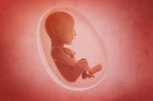
The amniotic sac, in which the fetus develops, consists of two membranes that are joined to one another. The amniotic membrane is in contact with the amniotic liquid and the fetus. The chorionic membrane, the outermost membrane, is in contact with the mother’s tissue. When the amniotic sac breaks (when a woman’s water breaks), these membranes tear, and the amniotic liquid comes out.
The maternal part of the amniotic sac contains chorionic villus. These are structures that are in contact with the mother’s endometrium, allowing for blood circulation between the mother and her developing baby.
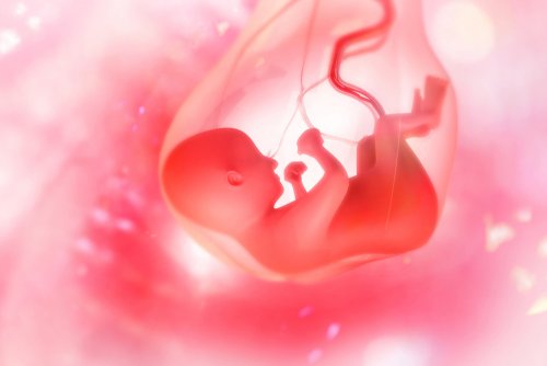
The umbilical cord, consisting of two arteries and a vein, connects the fetus with the maternal part of the placenta. Unlike adult circulation, in umbilical cord circulation, it’s the arteries that transport deoxygenated blood to the fetus to the placenta. At the same time, the vein is responsible for transporting oxygenated and nutrient-rich blood to the fetus.
In general, the placenta is located in the fundus (the top of the mother’s uterus). However, sometimes it can insert itself in other areas.
The functions of the placenta
The placenta has multiple functions that are fundamental for the proper development of the fetus:
- It allows for the exchange of gases and nutrients between mother and fetus.
- Hormone production: Chorionic gonadotropin, progesterone and placental lactogen.
- The placenta protects the fetus from maternal immune response, preventing the woman’s body from rejecting the fetus as a foreign body.
Each of the above-mentioned hormones has a specific function:
- Chorionic gonadotropin keeps the corpus luteum functional. The corpus luteum is responsible for producing progesterone and other hormones until the placenta is fully functional. This is the hormone that is detected in pregnancy tests.
- The corpus luteum releases progesterone until the second month of pregnancy. At this point, the placenta takes over the secretion of this hormone. Progesterone serves the synthesis of fetal corticoids and participates in the formation of decidual cells in the uterus. These cells are vital for the fetus’ nutrition.
- Placental lactogen stimulates the development and secretion of the mammary gland. It also stimulates the growth of fetal organs and the weight of the placenta.
The protective function of the placenta is paramount. The embryo is a foreign body and contains proteins (synthesized from the father’s genes) that are strange to the mother’s immune system. Therefore, the mother’s body could reject the fetus as a response from her immune system.
Thanks to the production of immunosuppressive and immunomodulating factors during early gestation, the placenta prevents this rejection from taking place.
Alteration in the placenta
Size anomalies
The placenta usually weighs, on average, about 1.5 pounds. Placentamegoly refers to cases in which the placenta is abnormally large (greater than 1/6 the size of the fetus). Placentas can also be abnormally small.
Morphological anomalies
Normal placentas are circular and disc-shaped in form. However, variations in their shape may occur, such as the following:
- Bilobed placenta. The placenta is divided into one or more lobes.
- Succenturiated lobe. In this case, one or more lobes exist separately from the placental disc, joined by vascular connections.
- Placenta spuria. This is similar to succentuariated lobe, but without the vascular connections.
- Circumvallate placenta. Here, a central depression appears on the fetal side of the placenta, surrounded by a whitish ring. The ring is a fold between both membranes.
Anomalies in uterine penetration
Abnormal insertion of the placenta takes place in the endometrium (known as decidua during pregnancy). There are several variations to this abnormal insertion, leading to various types of placenta.
- Placenta Accreta. The villus adheres to the myometrium (uterine muscle). It’s in contact with the muscle, but doesn’t invade it.
- Placenta Increta. Here, the villus penetrate the myometrium.
- In cases of Placenta percreta, the villus pass through the myometrium and reach the peritoneal serosa and may even penetrate the abdominal cavity and invade neighboring organs.
Implantation anomalies
Placenta previa
Placenta previa is a condition that occurs when the placenta covers the opening of the mother’s cervix. It can even come to block the internal cervical orifice, impeding vaginal birth. There are different grades of placenta previa:
- Minor (grade 1): Only a small part of the placenta extends to the lower part of the womb.
- Marginal (grade 2): The placenta reaches the cervix, but doesn’t cover it.
- Major (grade 3): The placenta covers the cervix, but only partially.
- Major (grade 4): The placenta covers the cervix completely. This is the most serious type of placenta previa.
Tumorous alterations
The following tumorous alterations are very uncommon, but may also occur.
- Hydatidiform Mole. This consists of the development of a mass located in the placenta. The tumor is non-invasive, neo-plastic and non-malignant. The mass develops after the implantation of the fertilized egg and, in general, in absence of a fetus. When this alteration occurs, there are usually high levels of the B-HCG hormone in the mother’s blood.
- Gestational trophoblastic tumors. These alterations are relatively rare, but are very aggressive and serious. They can be malignant or benign tumors, and develop from the placenta.
Vascular anomalies
Insufficient placenta can occur when the placenta doesn’t adequately perform its functions. This may be due to aging, placental infarction, among other factors.
One last note about the development of the placenta
If you have the opportunity, ask your gynecologist or the doula who accompanies you during childbirth to allow you to see your placenta. It’s an incredible organ that allows your baby to develop correctly. Its value is incalculable!
Learn about the different structures, functions and development of the placenta, which is an indispensable organ when it comes to fetal development during pregnancy.
This organ, that only appears when a woman is pregnant, allows for the nutrition and oxygenation of the fetus. The placenta also allows the fetus to eliminate its waste products. In this way, the placenta guarantees proper growth and well-being.
Development of the placenta
The development of the placenta goes through various phases during pregnancy. It begins on the 5th day after fertilization, in the preimplantation development stage, and continues throughout the rest of pregnancy. The organ produces changes until the very end of gestation.
What structures are present in the placenta?
The placenta is formed of two types of tissue: maternal and fetal. Fetal tissue includes the chorion, and maternal tissue includes the most superficial part of the uterine endometrium.
The amniotic sac, in which the fetus develops, consists of two membranes that are joined to one another. The amniotic membrane is in contact with the amniotic liquid and the fetus. The chorionic membrane, the outermost membrane, is in contact with the mother’s tissue. When the amniotic sac breaks (when a woman’s water breaks), these membranes tear, and the amniotic liquid comes out.
The maternal part of the amniotic sac contains chorionic villus. These are structures that are in contact with the mother’s endometrium, allowing for blood circulation between the mother and her developing baby.
The umbilical cord, consisting of two arteries and a vein, connects the fetus with the maternal part of the placenta. Unlike adult circulation, in umbilical cord circulation, it’s the arteries that transport deoxygenated blood to the fetus to the placenta. At the same time, the vein is responsible for transporting oxygenated and nutrient-rich blood to the fetus.
In general, the placenta is located in the fundus (the top of the mother’s uterus). However, sometimes it can insert itself in other areas.
The functions of the placenta
The placenta has multiple functions that are fundamental for the proper development of the fetus:
- It allows for the exchange of gases and nutrients between mother and fetus.
- Hormone production: Chorionic gonadotropin, progesterone and placental lactogen.
- The placenta protects the fetus from maternal immune response, preventing the woman’s body from rejecting the fetus as a foreign body.
Each of the above-mentioned hormones has a specific function:
- Chorionic gonadotropin keeps the corpus luteum functional. The corpus luteum is responsible for producing progesterone and other hormones until the placenta is fully functional. This is the hormone that is detected in pregnancy tests.
- The corpus luteum releases progesterone until the second month of pregnancy. At this point, the placenta takes over the secretion of this hormone. Progesterone serves the synthesis of fetal corticoids and participates in the formation of decidual cells in the uterus. These cells are vital for the fetus’ nutrition.
- Placental lactogen stimulates the development and secretion of the mammary gland. It also stimulates the growth of fetal organs and the weight of the placenta.
The protective function of the placenta is paramount. The embryo is a foreign body and contains proteins (synthesized from the father’s genes) that are strange to the mother’s immune system. Therefore, the mother’s body could reject the fetus as a response from her immune system.
Thanks to the production of immunosuppressive and immunomodulating factors during early gestation, the placenta prevents this rejection from taking place.
Alteration in the placenta
Size anomalies
The placenta usually weighs, on average, about 1.5 pounds. Placentamegoly refers to cases in which the placenta is abnormally large (greater than 1/6 the size of the fetus). Placentas can also be abnormally small.
Morphological anomalies
Normal placentas are circular and disc-shaped in form. However, variations in their shape may occur, such as the following:
- Bilobed placenta. The placenta is divided into one or more lobes.
- Succenturiated lobe. In this case, one or more lobes exist separately from the placental disc, joined by vascular connections.
- Placenta spuria. This is similar to succentuariated lobe, but without the vascular connections.
- Circumvallate placenta. Here, a central depression appears on the fetal side of the placenta, surrounded by a whitish ring. The ring is a fold between both membranes.
Anomalies in uterine penetration
Abnormal insertion of the placenta takes place in the endometrium (known as decidua during pregnancy). There are several variations to this abnormal insertion, leading to various types of placenta.
- Placenta Accreta. The villus adheres to the myometrium (uterine muscle). It’s in contact with the muscle, but doesn’t invade it.
- Placenta Increta. Here, the villus penetrate the myometrium.
- In cases of Placenta percreta, the villus pass through the myometrium and reach the peritoneal serosa and may even penetrate the abdominal cavity and invade neighboring organs.
Implantation anomalies
Placenta previa
Placenta previa is a condition that occurs when the placenta covers the opening of the mother’s cervix. It can even come to block the internal cervical orifice, impeding vaginal birth. There are different grades of placenta previa:
- Minor (grade 1): Only a small part of the placenta extends to the lower part of the womb.
- Marginal (grade 2): The placenta reaches the cervix, but doesn’t cover it.
- Major (grade 3): The placenta covers the cervix, but only partially.
- Major (grade 4): The placenta covers the cervix completely. This is the most serious type of placenta previa.
Tumorous alterations
The following tumorous alterations are very uncommon, but may also occur.
- Hydatidiform Mole. This consists of the development of a mass located in the placenta. The tumor is non-invasive, neo-plastic and non-malignant. The mass develops after the implantation of the fertilized egg and, in general, in absence of a fetus. When this alteration occurs, there are usually high levels of the B-HCG hormone in the mother’s blood.
- Gestational trophoblastic tumors. These alterations are relatively rare, but are very aggressive and serious. They can be malignant or benign tumors, and develop from the placenta.
Vascular anomalies
Insufficient placenta can occur when the placenta doesn’t adequately perform its functions. This may be due to aging, placental infarction, among other factors.
One last note about the development of the placenta
If you have the opportunity, ask your gynecologist or the doula who accompanies you during childbirth to allow you to see your placenta. It’s an incredible organ that allows your baby to develop correctly. Its value is incalculable!
All cited sources were thoroughly reviewed by our team to ensure their quality, reliability, currency, and validity. The bibliography of this article was considered reliable and of academic or scientific accuracy.
- Apaza Valencia John. (2014). Desarrollo placentario temprano: aspectos fisiopatológicos. Rev. peru. ginecol. obstet. 2014: 60( 2 ): 131-140. Disponible en: http://www.scielo.org.pe/scielo.php?script=sci_arttext&pid=S2304-51322014000200006&lng=es
- Roa Ignacio, Smok S Carolina, Prieto G Ruth. (2012). Placenta: Compared Anatomy and Histology. Int. J. Morphol. 2012: 30( 4 ): 1490-1496. Disponible en: https://scielo.conicyt.cl/scielo.php?script=sci_arttext&pid=S0717-95022012000400036&lng=es
- Espinilla Sanz, B., Tomé Blanco, E., Sadornil Vicario, M., Albillos Alonso, L. (2016). Alteraciones de la placenta. Manual de obstetricia para matronas. 2nd ed. Valladolid: DIFÁCIL; 2016. 185-188.
This text is provided for informational purposes only and does not replace consultation with a professional. If in doubt, consult your specialist.


