What Is a Chorionic Biopsy?

When an obstetrician determines that a current pregnancy is at high risk for genetic diseases, they may choose to perform different diagnostic studies. Among them, chorionic biopsy, which is a procedure that involves obtaining cells from a layer of the placenta: The chorion. It’s used to analyze the baby’s DNA, in order to diagnose possible alterations in its genes from the beginning of pregnancy.
Do you want to know more about what this procedure involves and what its implications are? Below, we’ll tell you everything you need to know. Keep reading!
What is a chorionic biopsy?
A chorionic biopsy is an invasive prenatal test, as it requires the collection of a sample of the placental chorionic villi through a surgical procedure.
The chorion is the outer membrane that surrounds the fetus within the uterus and has small villi or projections of placental tissue. Both this layer and the baby have the same genetic information.
From the villi sample, different aspects of this material can be analyzed.
Read more: Genetic Syndromes: What Are They and How Are They Inherited?
What does this procedure detect?
Next, we’ll tell you what specific information about the fetus doctors can obtain from the chorionic biopsy :
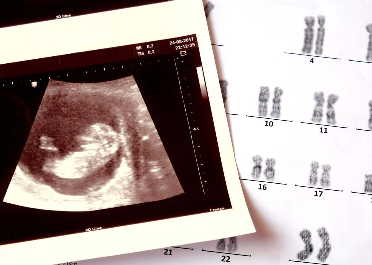
- Fetal karyotype: Each of the chromosomes that contain the genetic information is analyzed to find out the number and structure of each one.
- The sex of the baby: Thanks to the karyotype, it’s possible to determine the sex of the baby through the analysis of the sex chromosomes.
- Fetal metabolic disorders.
- Chromosomal abnormalities such as Down or Edwards syndrome) and genetic abnormalities (such as cystic fibrosis).
- Congenital infections (such as rubella, cytomegalovirus, toxoplasmosis, among others).
When does it take place?
A chorionic biopsy is a study that takes place during the first trimester of pregnancy, specifically between weeks 10 and 14.
It’s not a routine study for all pregnancies, but rather for situations where there may be a risk to the baby’s health, such as the following:
- A positive result of the first trimester screening
- History of chromosomal abnormalities in previous pregnancies
- Suspicion of malformations on the first trimester ultrasound
- Personal or family history of genetic diseases
- Elderly mothers
- History of repeated miscarriages
Likewise, doctors can perform a chorionic biopsy to confirm a preimplantation diagnosis in the case of certain assisted reproductive techniques.
Contraindications for chorionic biopsy
- Pregnancies of less than 10 weeks, due to the risk of causing defects in the extremities, such as shortening of the limbs
- Women who are seropositive for hepatitis B and C virus or human immunodeficiency virus with high viral load
- Women with isoimmunization
- The threat of abortion and bleeding
- Active maternal infection, with or without fever
- Intrauterine hematoma
- Maternal clotting problems
How is the chorionic biopsy performed?
This test requires an invasive technique, so a doctor must perform it in a germ-free area, such as the operating room.
It doesn’t require prior preparation and, in case of taking any medication, the obstetrician must indicate how to proceed with this.
This is a relatively short study, taking no more than 45 minutes.
To begin, the woman must put on a surgical gown and lie on the table in a comfortable position, which in turn facilitates the test. During the entire procedure, her vital signs are monitored and an ultrasound control is performed to evaluate the most appropriate site to take the sample.
There are two routes to obtain the villi: The transabdominal route and the transcervical route. It’s important to note that in neither case is the baby’s amniotic sac touched.
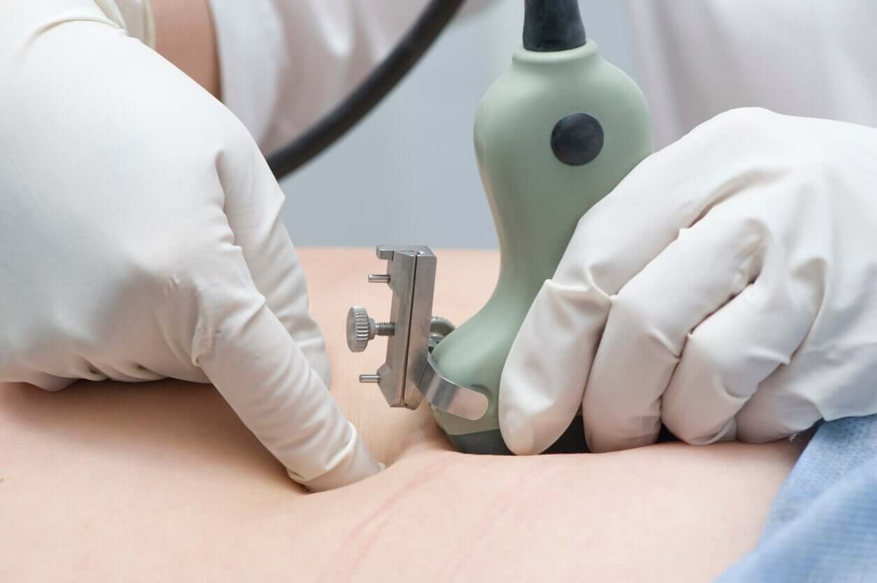
Transabdominal route
Access to the placenta is through the abdomen, with a fine needle that aspirates the necessary material. To avoid discomfort, doctors place local anesthesia on the mother’s skin and perform adequate asepsis. As we mentioned above, the puncture takes place under permanent ultrasound control.
Transcervical route
In this case, doctors access the placenta through the cervix. For this, they must perform proper asepsis of the intimate area, insert the speculum, and then, insert a semi-rigid fine forceps or cannula through the cervix.
Through this element, they take the chorionic villi sample, always with the guidance of ultrasound. This route is the most common because it allows for the taking of a greater amount of tissue.
Generally, it’s a painless procedure or with mild discomfort, similar to that of menstruation.
Some special considerations
If the mother is Rh-negative, she should receive an injection of anti-D immune globulin within 72 hours after the biopsy. This is because there’s the possibility that the baby’s blood will mix with that of its mother during the procedure.
At the same time, it’s important for the woman to maintain relative rest for 48 to 72 hours and avoid carrying weight, doing physical exercise, and having sexual intercourse.
Complications that may appear after chorionic biopsy
Although complications after the procedure are rare, there’s a minimal risk of developing them. Among these, the following stand out:
- Abdominal pain in the puncture area.
- Vaginal bleeding
- Infection of amniotic fluid and amniotic membrane (chorioamnionitis)
- Fever
- Premature rupture of membranes
- Placental hematoma
- Miscarriage (Less than 1%)
Nowadays, these complications have diminished considerably thanks to advances in techniques and equipment. However, any invasive procedure has its risks. The important thing is to go to the medical evaluation in case of feeling any discomfort.
When the benefits outweigh the risks
The chorionic biopsy is an accurate and reliable study, which provides a valid result in 99% of cases. For this reason, it’s the best choice to perform when doctors suspect potentially serious illnesses in the baby.
Preliminary results are available in 48 hours and the final result is ready in one week. The gynecologist and genetics will evaluate and deliver them. Only 2% of cases should require corroboration with amniocentesis.
Therefore, we can say that, in the face of any condition of potential risk, the benefits of doing this test are countless. Talk to your doctor and address any doubts you may have.
All cited sources were thoroughly reviewed by our team to ensure their quality, reliability, currency, and validity. The bibliography of this article was considered reliable and of academic or scientific accuracy.
- Bernal, L. (2017). Amniocentesis precoz y biopsia de vellosidad corial. Pérdidas fetales y anomalías congénitas en un grupo de gestantes brasileñas. Brasil. Recuperado de: http://www.scielo.org.co/pdf/nova/v16n29/1794-2470-nova-16-29-00051.pdf
- Garcia, R. (2011) Biopsia corial transcervical: guía práctica. España. Recuperado de: https://www.elsevier.es/es-revista-diagnostico-prenatal-327-articulo-biopsia-corial-transcervical-guia-practica-S2173412711001089
- Haumán, M. (2010). Procedimientos invasivos en el diagnóstico prenatal. Perú. Revista Peruana de Ginecología y Obstetricia. 2010; 56: 258-262
- Hospital Clinico Sant Joan de Deú. (2016) Protocolo: Biopsia de vellosidades coriales. España.