The Types of Ultrasounds in Pregnancy


Written and verified by the pediatrician Marcela Alejandra Caffulli
In the following article, we’re going to tell you all about the different types of ultrasounds during pregnancy and what each one is for. Join us!
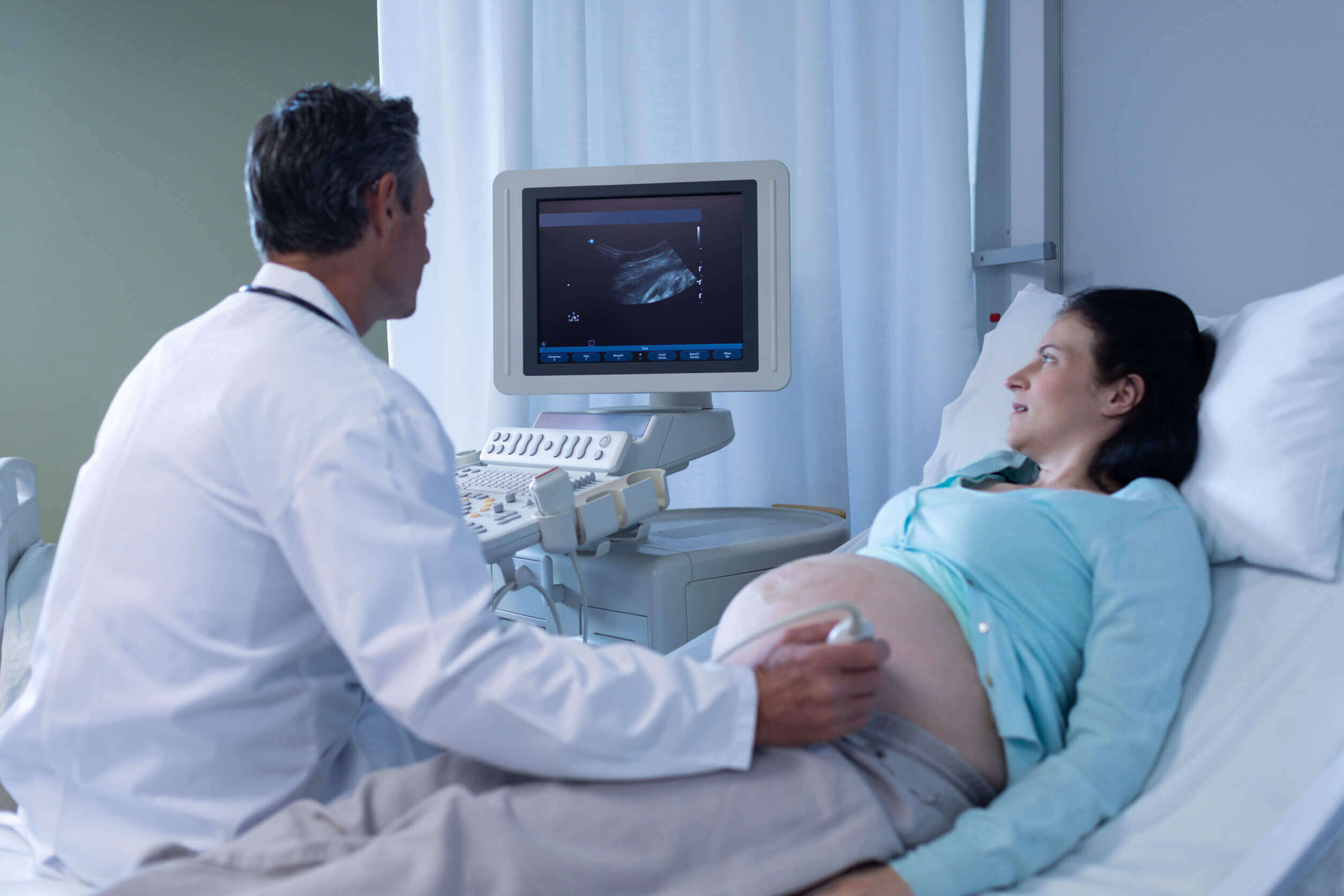
After seeing a positive pregnancy test, the first ultrasound is one of the most memorable moments for future parents. As the months go by, this study offers different possibilities of making contact with what happens inside the uterus.
Although ultrasounds are a safe study for the health of the mother and baby, experts recommend that they be used judiciously and only under precise medical indications. We mustn’t forget that they’re a diagnostic method and not a prenatal photography session.
Ultrasound of pregnancy: A window to observe the unborn baby
“Seeing is believing.” That phrase fits perfectly to describe the feeling of a mother seeing that she harbors a life inside her body.
Many women go through the first trimester without symptoms and some don’t even really take notice of their state until several months after conceiving. For this reason, obstetric ultrasound becomes a magic window in order to view that baby that lives inside the uterus.
Scientists have made amazing advances in this diagnostic method for more than half a century. In fact, it’s now possible to explore the maternal body during all stages of gestation. This study is indicated to know the status of the fetus, the placenta, the amniotic fluid, and the umbilical cord, among other issues.
“Ultrasound provides useful information, in real time, both for the diagnosis of obstetric disease and for adopting behaviors in daily clinical practice”.
-R González de Agüero Laborda-
How many types of pregnancy ultrasounds are there?
The different types of ultrasounds are done in much the same way: They require a special device that emits sound waves (an ultrasound machine), a computer that translates the echoes into images, and a doctor specially trained to practice and interpret what’s observed.
This last component is the determining factor for the quality of the method, which is why ultrasound is commonly defined as an “operator-dependent” study.
There are several ways to classify the different types of ultrasounds, either by the quality of the images obtained (in 2, 3, or 4 dimensions) or by the objectives set in each instance (standard screening, limited evaluation, and specialized evaluation).
1. Standard ultrasound evaluation
This is the type of ultrasound that’s practiced in almost all pregnancies, in order to know certain parameters of the baby and the attached organs (cord, placenta, fluids, among others).
During the first trimester, obstetric ultrasounds are indicated to determine the following:
- The presence of the embryo in the maternal uterus, the implantation site, and its size
- The number of gestational sacs and embryos in that cavity
- The approximate gestational age
- Embryonic cardiac activity, which can be recorded through two-dimensional (2D) techniques
- The state of the mother’s uterus, ovaries, and abdominal cavity
In the remaining two trimesters, routine ultrasounds examine the development of all these structures and some more aspects are added:
- Fetal growth (biometrics)
- Heart activity and other signs of vitality of the baby
- Screening for fetal abnormalities (morphological ultrasound)
- The volume of amniotic fluid (normo, poly, or oligohydramnios)
- The position of the baby in relation to the maternal pelvis (presentation)
- The location of the placenta
2. Limited ultrasound evaluation
This assessment is intended to monitor a specific aspect of maternal-fetal health that you wish to further explore.
In general, it’s performed after detecting a suspicious finding in a routine ultrasound, in order to confirm or rule out any disease or condition. They can include different techniques, such as Doppler.
3. Specialized ultrasound evaluation
This type of ultrasound is reserved for those situations in which there’s an increased risk of some type of fetal malformation. For example, when maternal age is considered advanced, in the event of an obstetric complication, or when conception occurred by assisted reproductive techniques.
Likewise, when there’s some degree of suspicion of fetal illness from laboratory studies or previous ultrasound scans.
These types of studies include the following ultrasound techniques:
- Fetal echocardiogram
- Fetal Doppler
- Cervical measurement through transvaginal ultrasound
- Nuchal translucency (NT): This study is performed between weeks 11 and 14 of gestation
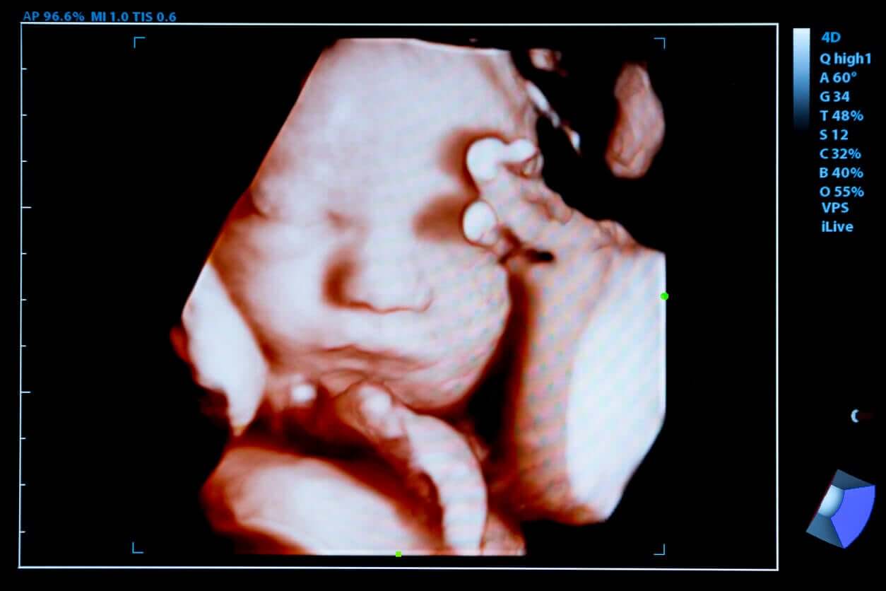
4. 3D and 4D ultrasounds
We mention these two types in a special section, due to the great popularity they’ve gained in recent years. But beyond the possibility they offer to see the baby’s face in detail, their real usefulness is debated by some specialists.
3 and 4-dimensional ultrasounds allow better visualization of the fetus and detect certain malformations, but this doesn’t imply that an early diagnosis of congenital syndromes can be made. More tools are needed to corroborate certain findings and this issue isn’t so straightforward.
Generally speaking, 3D ultrasound allows you to obtain a three-dimensional image of the fetus and 4D adds movement to it. Through the latter, it’s possible to see the child live as it moves about inside the mother’s womb.
Ultrasound Safety
For many years, the safety of the use of ultrasound in pregnant women has been questioned, but the different scientific societies of the world have concluded that it is. However, this study should only be carried out when medically indicated and appropriately. That is, with the necessary equipment and the suitable personnel for this purpose.
One of the main potential risks attributed to ultrasound studies was thermal injury. At present, the configuration of the equipment for obstetric studies avoids the increase of the temperature of the maternal-fetal tissues.
In this sense, the American Institute of Ultrasound in Medicine (AIUM) suggests that when the study is carried out under the “ALARA” principles (As Low As Reasonably Achievable) the risk of causing harm to the baby is minimal.
Regarding the different types of ultrasounds, Doppler is the one that can generate the highest temperature within the uterine cavity. For this reason, it should be requested only when strictly necessary and used for the shortest possible time.
Talk to your doctor before having a pregnancy ultrasound!
As we¿ve said, ultrasounds allow us to know various aspects of the unborn baby and this has its benefits, but also its risks.
Although it’s a study safe enough to be performed during pregnancy, it must be used judiciously. In this sense, it shouldn’t be considered for social or entertainment purposes.
In the following article, we’re going to tell you all about the different types of ultrasounds during pregnancy and what each one is for. Join us!
After seeing a positive pregnancy test, the first ultrasound is one of the most memorable moments for future parents. As the months go by, this study offers different possibilities of making contact with what happens inside the uterus.
Although ultrasounds are a safe study for the health of the mother and baby, experts recommend that they be used judiciously and only under precise medical indications. We mustn’t forget that they’re a diagnostic method and not a prenatal photography session.
Ultrasound of pregnancy: A window to observe the unborn baby
“Seeing is believing.” That phrase fits perfectly to describe the feeling of a mother seeing that she harbors a life inside her body.
Many women go through the first trimester without symptoms and some don’t even really take notice of their state until several months after conceiving. For this reason, obstetric ultrasound becomes a magic window in order to view that baby that lives inside the uterus.
Scientists have made amazing advances in this diagnostic method for more than half a century. In fact, it’s now possible to explore the maternal body during all stages of gestation. This study is indicated to know the status of the fetus, the placenta, the amniotic fluid, and the umbilical cord, among other issues.
“Ultrasound provides useful information, in real time, both for the diagnosis of obstetric disease and for adopting behaviors in daily clinical practice”.
-R González de Agüero Laborda-
How many types of pregnancy ultrasounds are there?
The different types of ultrasounds are done in much the same way: They require a special device that emits sound waves (an ultrasound machine), a computer that translates the echoes into images, and a doctor specially trained to practice and interpret what’s observed.
This last component is the determining factor for the quality of the method, which is why ultrasound is commonly defined as an “operator-dependent” study.
There are several ways to classify the different types of ultrasounds, either by the quality of the images obtained (in 2, 3, or 4 dimensions) or by the objectives set in each instance (standard screening, limited evaluation, and specialized evaluation).
1. Standard ultrasound evaluation
This is the type of ultrasound that’s practiced in almost all pregnancies, in order to know certain parameters of the baby and the attached organs (cord, placenta, fluids, among others).
During the first trimester, obstetric ultrasounds are indicated to determine the following:
- The presence of the embryo in the maternal uterus, the implantation site, and its size
- The number of gestational sacs and embryos in that cavity
- The approximate gestational age
- Embryonic cardiac activity, which can be recorded through two-dimensional (2D) techniques
- The state of the mother’s uterus, ovaries, and abdominal cavity
In the remaining two trimesters, routine ultrasounds examine the development of all these structures and some more aspects are added:
- Fetal growth (biometrics)
- Heart activity and other signs of vitality of the baby
- Screening for fetal abnormalities (morphological ultrasound)
- The volume of amniotic fluid (normo, poly, or oligohydramnios)
- The position of the baby in relation to the maternal pelvis (presentation)
- The location of the placenta
2. Limited ultrasound evaluation
This assessment is intended to monitor a specific aspect of maternal-fetal health that you wish to further explore.
In general, it’s performed after detecting a suspicious finding in a routine ultrasound, in order to confirm or rule out any disease or condition. They can include different techniques, such as Doppler.
3. Specialized ultrasound evaluation
This type of ultrasound is reserved for those situations in which there’s an increased risk of some type of fetal malformation. For example, when maternal age is considered advanced, in the event of an obstetric complication, or when conception occurred by assisted reproductive techniques.
Likewise, when there’s some degree of suspicion of fetal illness from laboratory studies or previous ultrasound scans.
These types of studies include the following ultrasound techniques:
- Fetal echocardiogram
- Fetal Doppler
- Cervical measurement through transvaginal ultrasound
- Nuchal translucency (NT): This study is performed between weeks 11 and 14 of gestation
4. 3D and 4D ultrasounds
We mention these two types in a special section, due to the great popularity they’ve gained in recent years. But beyond the possibility they offer to see the baby’s face in detail, their real usefulness is debated by some specialists.
3 and 4-dimensional ultrasounds allow better visualization of the fetus and detect certain malformations, but this doesn’t imply that an early diagnosis of congenital syndromes can be made. More tools are needed to corroborate certain findings and this issue isn’t so straightforward.
Generally speaking, 3D ultrasound allows you to obtain a three-dimensional image of the fetus and 4D adds movement to it. Through the latter, it’s possible to see the child live as it moves about inside the mother’s womb.
Ultrasound Safety
For many years, the safety of the use of ultrasound in pregnant women has been questioned, but the different scientific societies of the world have concluded that it is. However, this study should only be carried out when medically indicated and appropriately. That is, with the necessary equipment and the suitable personnel for this purpose.
One of the main potential risks attributed to ultrasound studies was thermal injury. At present, the configuration of the equipment for obstetric studies avoids the increase of the temperature of the maternal-fetal tissues.
In this sense, the American Institute of Ultrasound in Medicine (AIUM) suggests that when the study is carried out under the “ALARA” principles (As Low As Reasonably Achievable) the risk of causing harm to the baby is minimal.
Regarding the different types of ultrasounds, Doppler is the one that can generate the highest temperature within the uterine cavity. For this reason, it should be requested only when strictly necessary and used for the shortest possible time.
Talk to your doctor before having a pregnancy ultrasound!
As we¿ve said, ultrasounds allow us to know various aspects of the unborn baby and this has its benefits, but also its risks.
Although it’s a study safe enough to be performed during pregnancy, it must be used judiciously. In this sense, it shouldn’t be considered for social or entertainment purposes.
All cited sources were thoroughly reviewed by our team to ensure their quality, reliability, currency, and validity. The bibliography of this article was considered reliable and of academic or scientific accuracy.
- Callen P, Norton ME. Ecografía Obstétrica. Capítulo 1. En: Norton ME, Scout LM, Feldstein VA. Callen Ecografía en Obstertricia y Ginecología. Sexta edición. Elsevier España, 2018.
- Committee on Obstetric Practice, American College of Obstetricians and Gynecologists (ACOG). Guidelines for Diagnostic Imaging During Pregnancy and Lactation. Number 723, October 2017. Reaffirmed October 2021. Disponible en: https://www.acog.org/clinical/clinical-guidance/committee-opinion/articles/2017/10/guidelines-for-diagnostic-imaging-during-pregnancy-and-lactation
- American Institute of Ultrasound in Medicine: AIUM practice guideline for the performance of obstetric ultrasound examinations. J Ultrasound Med 2018; 9999:1-12. Disponible en: https://www.aium.org/resources/guidelines/obstetric.pdf
- González de Agüero Laborda R, Sobreviela Laserrada M, Espinosa A, Fabre González E. Diagnóstico por la imagen en el embarazo. Capítulo 9. En: González-Merlo. Obstetricia. Séptima edición. Elsevier España, 2018. Páginas 127-157
- American Institute of Ultrasound in Medicine (AIUM). Statement on the safe use of Doppler ultrasound during 11-14 week scans (or earlier in pregnancy) . Laurel (MD): AIUM; 2011. Disponible en: http://www.aium.org/officialStatements/42.
- Barišić L.S., Stanojević M., Kurjak A., Porović S., Gaber G.: Diagnosis of fetal syndromes by three- and four-dimensional ultrasound: is there any improvement?. J Perinat Med 2017; 45: pp. 651-665. Disponible en: https://pubmed.ncbi.nlm.nih.gov/28493822/
This text is provided for informational purposes only and does not replace consultation with a professional. If in doubt, consult your specialist.


