Third-Trimester Ultrasound: What You Need to Know


Written and verified by the nurse Leidy Mora Molina
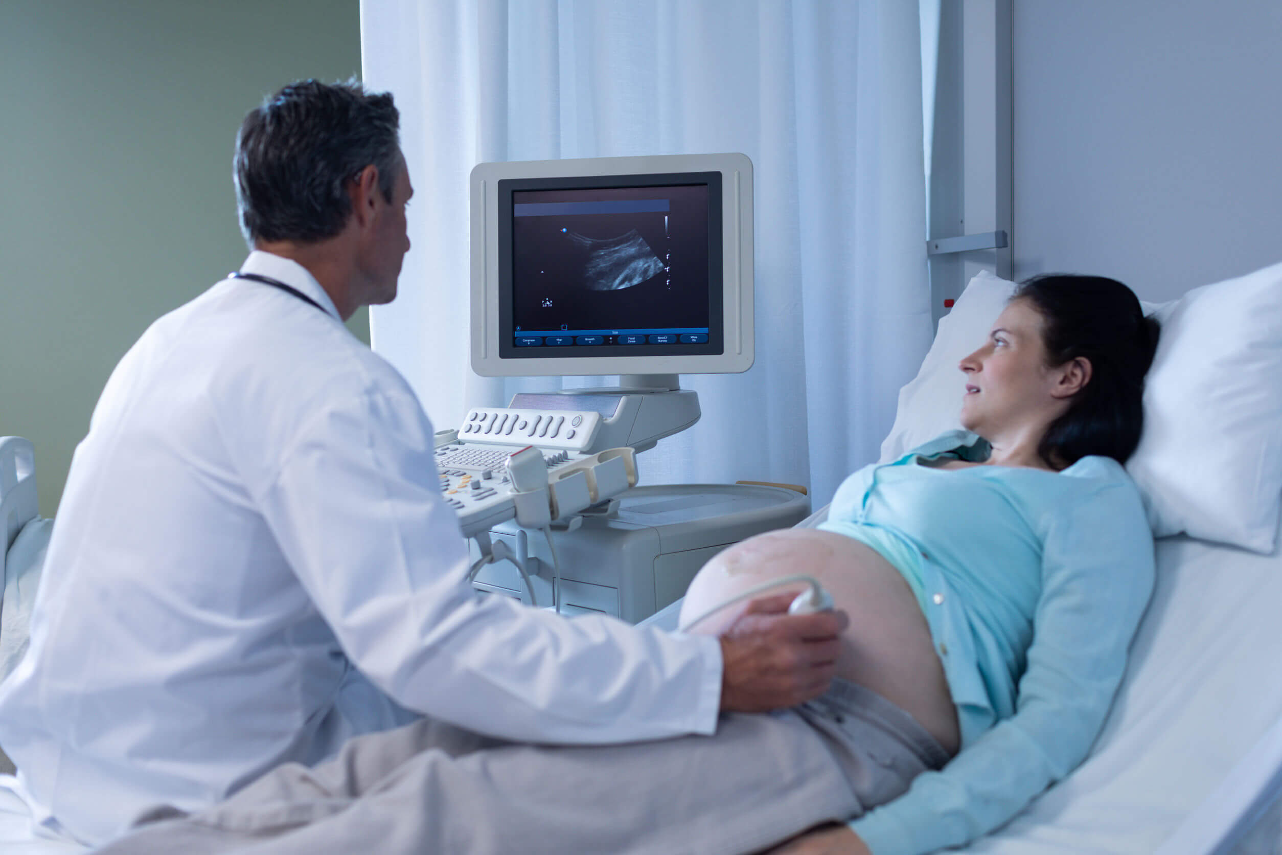
Throughout pregnancy, doctors will order at least 3 ultrasounds to assess the baby’s growth and development, as well as the condition of the mother’s uterine tissues. The third-trimester ultrasound is usually the last one to be performed and is focused on corroborating fetal wellbeing in the final stretch.
Find out what other parameters are assessed in this study, in which week it’s performed, and why it’s key that you have it done. Keep reading!
In what week is the third-trimester ultrasound performed?
As we always emphasize, ultrasounds are a non-invasive, fast, simple, and painless diagnostic study. They’re used to observe the state of the internal structures through the emission of radiofrequency waves.
As for prenatal ultrasounds, the main advantage they offer is that they allow the visual monitoring of the pregnancy in order to detect certain gestational complications in a timely manner.
Throughout the 9 months of gestation, gynecologists indicate several ultrasounds to verify the correct growth and development of the baby, as well as the adaptation of the maternal tissues to this new state.
Although all ultrasounds are important, the one performed in the third trimester is essential to anticipate behaviors related to the birth of the baby. For this reason, it’s usually performed between the 32nd and 36th week of pregnancy.

What is assessed in the third-trimester ultrasound?
The third-trimester ultrasound is aimed at observing the following parameters of maternal and fetal health:
- Fetal growth: the last stage of pregnancy is characterized by the “fattening” of the fetus. Therefore, one of the main data to be collected with this study is to know if the size of the fetus is in accordance with its gestational age. When it’s smaller than expected, it’s necessary to complete the evaluation with other studies in order to rule out intrauterine growth retardation.
- Biophysical profile of the baby: in addition to size, the physician assesses heart rate, respiratory movements, activity, and fetal tone. All this information is very relevant to assess whether the baby is ready to leave the uterus and whether the conditions inside the uterus are adequate.
- Fetal statics: that is, the relative position of the baby inside the uterus. Whether it is head first (cephalic presentation), breech, or transverse.
- Fetal anatomy: although this point is evaluated in detail in the morphological ultrasound, it’s re-evaluated in this instance in order to detect late-onset congenital anomalies.
- Placental status: in addition to corroborating the baby’s health, the degree of placental maturity is also determined. When the placenta is aged or at risk of premature aging, it’s best to closely monitor the pregnancy. Also, the location of the placenta is corroborated and the most appropriate route of birth is defined in the case of placenta previa.
- Amount of amniotic fluid: this parameter reports the volume of fluid surrounding the baby, which must be kept within strict margins to avoid gestational complications.
- Functionality of the umbilical cord: this structure guarantees the arrival of oxygen and nutrients to the baby, so in each ultrasound, its anatomy and position are evaluated and cord knots are ruled out. To complete the evaluation, the physician may request a Doppler ultrasound to assess the blood flow through this structure.
- Cervical length: this consists of measuring the length of the cervix, in order to rule out the risk of premature births.
- Position of the fetuses in a twin pregnancy: the position adopted by the two or more babies in a multiple pregnancy will make it possible to define the most appropriate route of delivery.
Other parameters that can be assessed in this ultrasound scan
It’s possible that in this encounter, a visualization of the baby with the 4D technique is performed. This way, the physician will be able to detail the facial features with greater precision, as well as the cardiac and cerebral anatomy of the little one.
As for the mother, in the case of pelvic tumors, such as large myomas, this ultrasound is the last evaluation. The location and size acquired during gestation are defined and the most convenient route of delivery is evaluated.
How is the third trimester ultrasound performed?
No preparation is required for the third-trimester ultrasound. However, it’s advisable to go to the clinic with loose and comfortable clothing, which facilitates the exposure of the belly. Also, it’s best not to put moisturizing creams on the skin of the belly, so as not to interfere with the ultrasound image capture.
This study is performed through the abdominal route, so it is necessary to empty the bladder before starting to facilitate the visualization. In general, it usually lasts between 15 and 30 minutes.
Keep in mind that due to the size of the baby, only specific areas of its body can be observed. On the other hand, if the specialist wishes to carefully evaluate the length of the cervix or the characteristics of the placenta, it’s possible to complement the study with a transvaginal ultrasound.
Don’t miss any prenatal studies
Prenatal ultrasounds are a practical method to assess the welfare of your baby throughout pregnancy and don’t represent any danger to health. Generally, one per trimester is indicated, but this will depend on the findings or the mother’s underlying conditions.
Take advantage of all the benefits of ultrasounds and don’t miss any prenatal study. This way, you’ll provide the best care for your child from the very first moment.
Throughout pregnancy, doctors will order at least 3 ultrasounds to assess the baby’s growth and development, as well as the condition of the mother’s uterine tissues. The third-trimester ultrasound is usually the last one to be performed and is focused on corroborating fetal wellbeing in the final stretch.
Find out what other parameters are assessed in this study, in which week it’s performed, and why it’s key that you have it done. Keep reading!
In what week is the third-trimester ultrasound performed?
As we always emphasize, ultrasounds are a non-invasive, fast, simple, and painless diagnostic study. They’re used to observe the state of the internal structures through the emission of radiofrequency waves.
As for prenatal ultrasounds, the main advantage they offer is that they allow the visual monitoring of the pregnancy in order to detect certain gestational complications in a timely manner.
Throughout the 9 months of gestation, gynecologists indicate several ultrasounds to verify the correct growth and development of the baby, as well as the adaptation of the maternal tissues to this new state.
Although all ultrasounds are important, the one performed in the third trimester is essential to anticipate behaviors related to the birth of the baby. For this reason, it’s usually performed between the 32nd and 36th week of pregnancy.
What is assessed in the third-trimester ultrasound?
The third-trimester ultrasound is aimed at observing the following parameters of maternal and fetal health:
- Fetal growth: the last stage of pregnancy is characterized by the “fattening” of the fetus. Therefore, one of the main data to be collected with this study is to know if the size of the fetus is in accordance with its gestational age. When it’s smaller than expected, it’s necessary to complete the evaluation with other studies in order to rule out intrauterine growth retardation.
- Biophysical profile of the baby: in addition to size, the physician assesses heart rate, respiratory movements, activity, and fetal tone. All this information is very relevant to assess whether the baby is ready to leave the uterus and whether the conditions inside the uterus are adequate.
- Fetal statics: that is, the relative position of the baby inside the uterus. Whether it is head first (cephalic presentation), breech, or transverse.
- Fetal anatomy: although this point is evaluated in detail in the morphological ultrasound, it’s re-evaluated in this instance in order to detect late-onset congenital anomalies.
- Placental status: in addition to corroborating the baby’s health, the degree of placental maturity is also determined. When the placenta is aged or at risk of premature aging, it’s best to closely monitor the pregnancy. Also, the location of the placenta is corroborated and the most appropriate route of birth is defined in the case of placenta previa.
- Amount of amniotic fluid: this parameter reports the volume of fluid surrounding the baby, which must be kept within strict margins to avoid gestational complications.
- Functionality of the umbilical cord: this structure guarantees the arrival of oxygen and nutrients to the baby, so in each ultrasound, its anatomy and position are evaluated and cord knots are ruled out. To complete the evaluation, the physician may request a Doppler ultrasound to assess the blood flow through this structure.
- Cervical length: this consists of measuring the length of the cervix, in order to rule out the risk of premature births.
- Position of the fetuses in a twin pregnancy: the position adopted by the two or more babies in a multiple pregnancy will make it possible to define the most appropriate route of delivery.
Other parameters that can be assessed in this ultrasound scan
It’s possible that in this encounter, a visualization of the baby with the 4D technique is performed. This way, the physician will be able to detail the facial features with greater precision, as well as the cardiac and cerebral anatomy of the little one.
As for the mother, in the case of pelvic tumors, such as large myomas, this ultrasound is the last evaluation. The location and size acquired during gestation are defined and the most convenient route of delivery is evaluated.
How is the third trimester ultrasound performed?
No preparation is required for the third-trimester ultrasound. However, it’s advisable to go to the clinic with loose and comfortable clothing, which facilitates the exposure of the belly. Also, it’s best not to put moisturizing creams on the skin of the belly, so as not to interfere with the ultrasound image capture.
This study is performed through the abdominal route, so it is necessary to empty the bladder before starting to facilitate the visualization. In general, it usually lasts between 15 and 30 minutes.
Keep in mind that due to the size of the baby, only specific areas of its body can be observed. On the other hand, if the specialist wishes to carefully evaluate the length of the cervix or the characteristics of the placenta, it’s possible to complement the study with a transvaginal ultrasound.
Don’t miss any prenatal studies
Prenatal ultrasounds are a practical method to assess the welfare of your baby throughout pregnancy and don’t represent any danger to health. Generally, one per trimester is indicated, but this will depend on the findings or the mother’s underlying conditions.
Take advantage of all the benefits of ultrasounds and don’t miss any prenatal study. This way, you’ll provide the best care for your child from the very first moment.
All cited sources were thoroughly reviewed by our team to ensure their quality, reliability, currency, and validity. The bibliography of this article was considered reliable and of academic or scientific accuracy.
- Gonzales, A. (2009). Ecografía en obstetricia. Anales de Pediatría Continuada. Vol. 7. Núm. 1. Recuperado de: https://www.elsevier.es/es-revista-anales-pediatria-continuada-51-articulo-ecografia-obstetricia-S1696281809704500
- Marcus, G. Et al (2020). Características del circular de cordón umbilical simple en neonatos de partos eutócicos atendidos en servicio de primer nivel. Revista Científica de Salud UNITEPC. Vol.7 N°2. Recuperado de: http://www.scielo.org.bo/scielo.php?pid=S2520-98252020000200002&script=sci_arttext
- Vila-Candel, R. et al (2019). Ecografía del tercer trimestre combinada con un método clínico para mejorar la predicción del peso del recién nacido a término: un estudio de cohortes en España. Revista Colombiana de Obstetricia y Ginecología. Vol.70, Núm.1, pp.27-38. Recuperado de: http://www.scielo.org.co/scielo.php?script=sci_abstract&pid=S0034-74342019000100027&lng=en&nrm=iso&tlng=es
This text is provided for informational purposes only and does not replace consultation with a professional. If in doubt, consult your specialist.


