20-Week Ultrasound: Why is it So Important?

The 20-week ultrasound is a routine test that allows a thorough evaluation of the baby’s organs, the mother’s uterus, and the placenta. In addition to the valuable information it offers, it’s carried out at a key moment in pregnancy.
Do you know why it’s so important to have it done? Get to know the answers in this article.
Why do doctors order a 20-week ultrasound?
When you reach the middle of the pregnancy, your obstetrician usually requests a morphological ultrasound that, as its name indicates, seeks to analyze the baby’s anatomy.
This study is performed between weeks 18 and 20 of gestation for a number of reasons that favor fetal visualization:
- The amount of amniotic fluid is adequate to obtain clear images.
- The baby has acquired just the right size to examine its structures and also to see its entire body at once.
- The fetal organs are almost fully formed and their morphology and location can be thoroughly analyzed.
This way, the doctor can evaluate how the baby grows and develops inside the womb. And at the same time, the 20-week ultrasound allows for the early detection of any type of anomaly in any of these aspects.

What does the procedure consist of?
The 20-week ultrasound is one of the most comprehensive pregnancy tests, but it’s not too different from the ones you’ve had before. It doesn’t radiate or cause pain and it doesn’t cause pain or represent a risk to your baby’s health.
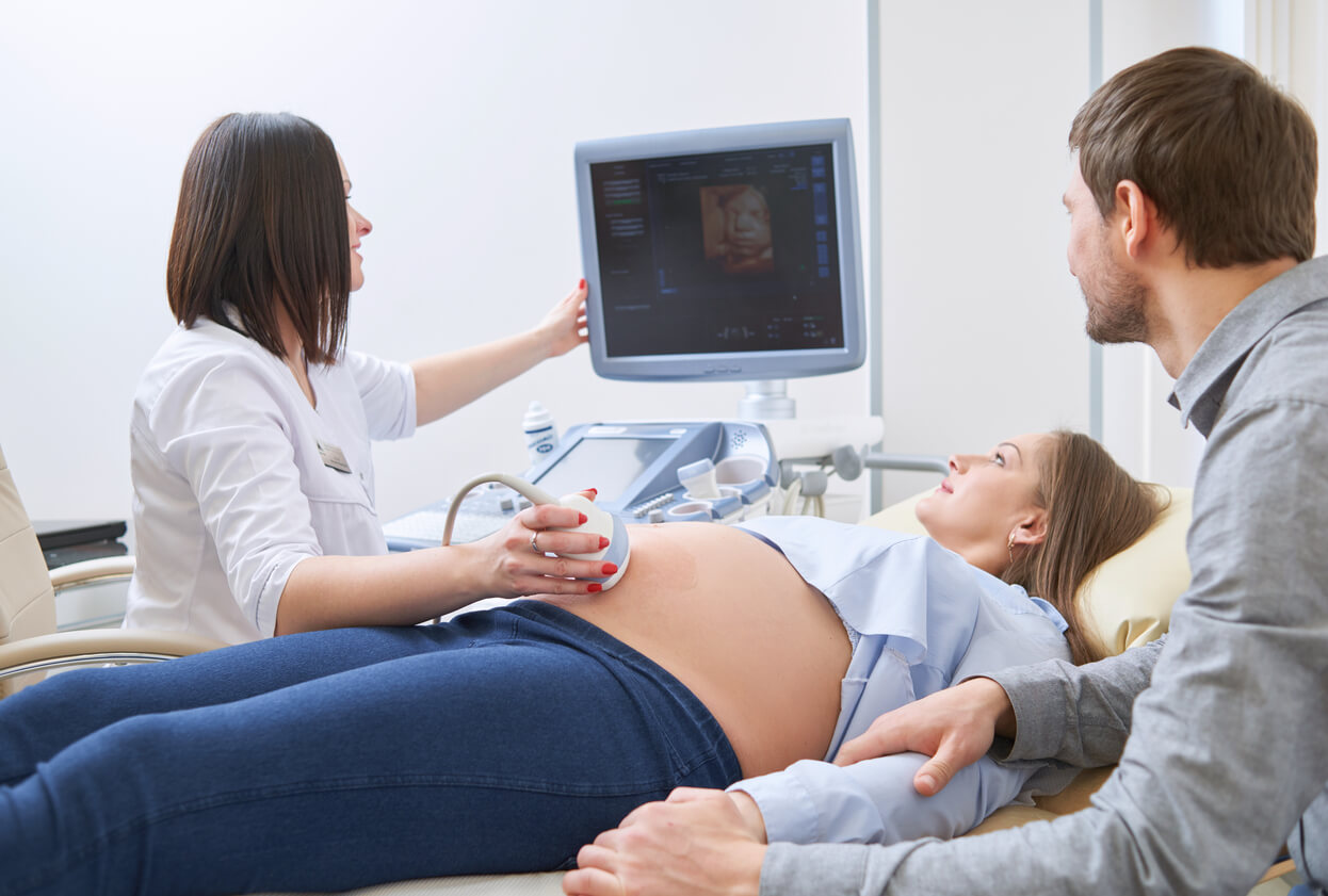
The 20-week ultrasound is performed abdominally and doesn’t require any special preparation. Once inside the office, the specialist will ask you to lie down on the stretcher and will place a gel on your abdomen. In general, the procedure takes about 30 minutes.
Although it’s not an invasive study, the doctor will provide you with some explanations about the state of your baby’s health, so you should be accompanied by your partner or a close relative. Among other things, it’s possible for the specialist to confirm the sex of the baby, in case you don’t already know it.
What information do doctors seek to obtain with this test?
As we’ve anticipated, the 20-week ultrasound seeks to analyze the morphology of the baby in detail to detect any problem in its organs and tissues as soon as possible. In addition, it corroborates the state of the placenta, the umbilical cord, and the maternal genital organs.
Generally speaking, the 20-week ultrasound is performed for the following purposes:
- To estimate the fetal biometry, that is, the measurements of the baby, and compare them with the gestational age. This allows for the detection of growth problems, such as intrauterine growth retardation.
- To assess fetal vitality by observing the baby’s tone and body movements.
- To check the blood flow that reaches the placenta and the baby (through the cord).
- To detect fetal abnormalities.
- And to evaluate uterine structures, such as the location and morphology of the placenta and the amount of amniotic fluid that’s present.
Is the 20-week ultrasound 3D?
In general, this ultrasound is 2D. However, if the device allows it, some images can be captured in 3D or 4D. The latter shows the baby’s facial features with greater precision and even shows their movements in real-time.
The specialist may combine 2D and 3D images for a more complete analysis of all of the baby’s structures.
What’s the importance of performing the 20-week ultrasound?
With the 20-week ultrasound, it’s possible to assess whether fetal growth and development are adequate for gestational age.
Also, specialists can detect the presence of congenital malformations or risk conditions of developing gestational complications. For example, those related to a low level of amniotic fluid or the position of the placenta.
In case the specialist detects a problem in the baby or in the maternal structures, they’ll indicate some more precise tests to corroborate the findings found.
It’s important to emphasize that the 20-week ultrasound doesn’t definitively exclude the presence of diseases in the baby. Although it’s a fairly accurate diagnostic test, there are some conditions that can’t be detected through this study.
At the same time, if there are technical problems when performing this ultrasound, such as an inadequate fetal position, a shortage of amniotic fluid, or a large adiposity in the mother’s abdomen, the specialist may decide to repeat it.
Make your appointment for the 20-week ultrasound
Before doing the 20-week ultrasound, it’s best to clarify all your doubts with your doctor. Sometimes medical terms can be a bit confusing and the images you see aren’t entirely clear to untrained eyes. Therefore, to avoid going home with unnecessary anguish, discuss everything with your trusted doctor.
The 20-week ultrasound is a routine test that allows a thorough evaluation of the baby’s organs, the mother’s uterus, and the placenta. In addition to the valuable information it offers, it’s carried out at a key moment in pregnancy.
Do you know why it’s so important to have it done? Get to know the answers in this article.
Why do doctors order a 20-week ultrasound?
When you reach the middle of the pregnancy, your obstetrician usually requests a morphological ultrasound that, as its name indicates, seeks to analyze the baby’s anatomy.
This study is performed between weeks 18 and 20 of gestation for a number of reasons that favor fetal visualization:
- The amount of amniotic fluid is adequate to obtain clear images.
- The baby has acquired just the right size to examine its structures and also to see its entire body at once.
- The fetal organs are almost fully formed and their morphology and location can be thoroughly analyzed.
This way, the doctor can evaluate how the baby grows and develops inside the womb. And at the same time, the 20-week ultrasound allows for the early detection of any type of anomaly in any of these aspects.
What does the procedure consist of?
The 20-week ultrasound is one of the most comprehensive pregnancy tests, but it’s not too different from the ones you’ve had before. It doesn’t radiate or cause pain and it doesn’t cause pain or represent a risk to your baby’s health.
The 20-week ultrasound is performed abdominally and doesn’t require any special preparation. Once inside the office, the specialist will ask you to lie down on the stretcher and will place a gel on your abdomen. In general, the procedure takes about 30 minutes.
Although it’s not an invasive study, the doctor will provide you with some explanations about the state of your baby’s health, so you should be accompanied by your partner or a close relative. Among other things, it’s possible for the specialist to confirm the sex of the baby, in case you don’t already know it.
What information do doctors seek to obtain with this test?
As we’ve anticipated, the 20-week ultrasound seeks to analyze the morphology of the baby in detail to detect any problem in its organs and tissues as soon as possible. In addition, it corroborates the state of the placenta, the umbilical cord, and the maternal genital organs.
Generally speaking, the 20-week ultrasound is performed for the following purposes:
- To estimate the fetal biometry, that is, the measurements of the baby, and compare them with the gestational age. This allows for the detection of growth problems, such as intrauterine growth retardation.
- To assess fetal vitality by observing the baby’s tone and body movements.
- To check the blood flow that reaches the placenta and the baby (through the cord).
- To detect fetal abnormalities.
- And to evaluate uterine structures, such as the location and morphology of the placenta and the amount of amniotic fluid that’s present.
Is the 20-week ultrasound 3D?
In general, this ultrasound is 2D. However, if the device allows it, some images can be captured in 3D or 4D. The latter shows the baby’s facial features with greater precision and even shows their movements in real-time.
The specialist may combine 2D and 3D images for a more complete analysis of all of the baby’s structures.
What’s the importance of performing the 20-week ultrasound?
With the 20-week ultrasound, it’s possible to assess whether fetal growth and development are adequate for gestational age.
Also, specialists can detect the presence of congenital malformations or risk conditions of developing gestational complications. For example, those related to a low level of amniotic fluid or the position of the placenta.
In case the specialist detects a problem in the baby or in the maternal structures, they’ll indicate some more precise tests to corroborate the findings found.
It’s important to emphasize that the 20-week ultrasound doesn’t definitively exclude the presence of diseases in the baby. Although it’s a fairly accurate diagnostic test, there are some conditions that can’t be detected through this study.
At the same time, if there are technical problems when performing this ultrasound, such as an inadequate fetal position, a shortage of amniotic fluid, or a large adiposity in the mother’s abdomen, the specialist may decide to repeat it.
Make your appointment for the 20-week ultrasound
Before doing the 20-week ultrasound, it’s best to clarify all your doubts with your doctor. Sometimes medical terms can be a bit confusing and the images you see aren’t entirely clear to untrained eyes. Therefore, to avoid going home with unnecessary anguish, discuss everything with your trusted doctor.
All cited sources were thoroughly reviewed by our team to ensure their quality, reliability, currency, and validity. The bibliography of this article was considered reliable and of academic or scientific accuracy.
- Gonzales, A, et al. (2009). Ecografía en obstetricia. Anales de Pediatría Continuada Vol. 7 Nº1.
- Organización mundial de la salud (2021) Anomalías congénitas. Recuperado de: https://www.who.int/es/news-room/fact-sheets/detail/congenital-anomalies
- Sociedad Argentina de Ultrasonografía en medicina y biología (2016) Guía para el estudio morfológico del segundo trimestre del embarazo. Argentina. Revista Argentina de Ultrasonido – 2016; Volumen 15 N°1: 372 – 379.
- Sociedad española de ginecología y obstetricia (2020). Guía sistemática de la exploración ecográfica del segundo trimestre. Progresos de Obstetricia y Ginecología 2020; Vol. 63 Nº 2:99-122
This text is provided for informational purposes only and does not replace consultation with a professional. If in doubt, consult your specialist.


